Recognition of blast cells with monocytic differentiation: Difference between revisions
From haematologyetc.co.uk
No edit summary |
No edit summary |
||
| Line 2: | Line 2: | ||
<div style="width: 200px"> | <div style="width: 200px"> | ||
{| class="wikitable" style="border-left:solid 5px green;border-right:solid 5px green;border-top:solid 5px black;border-bottom:solid 5px black; font-size:90%; color:navy" | {| class="wikitable" style="border-left:solid 5px green;border-right:solid 5px green;border-top:solid 5px black;border-bottom:solid 5px black; font-size:90%; color:navy" | ||
| colspan="1"''|[[ | | colspan="1"''|[[Atypical patterns of primitive marker expression in acute myeloid leukaemia|Return to previous page]]'' | ||
|} | |} | ||
</div> | </div> | ||
Revision as of 00:11, 12 January 2024
Recognition of blast cells with monocytic differentiation
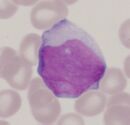
 A monoblastic AML
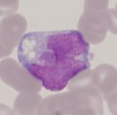
 B monocytic AML
- Panel A: Where cells have a monoblastic differentiation (generally large blast cells with primitive chromatin and nucleoli that lack monocytic shape often with basophilic cytoplasm) the monoblasts tend to express markers of primitive phenotype, while typical antigens of monocytic differentiation may be variable or weakly expressed.
- Panel B: Where cells undergo more appreciable monocytic differentiation (note the more condensed chromatin, lack of nucleolus, and monocytic shape of there nucleus) these cells tend to have stronger CD45 expression and often express typical monocyte antigens, primitive markers may be variably expressed or absent.
- Mixed patterns of monoblastic monocytic and myeloid differentiation within the same case are also frequently found.
- Correlation of immunophenotype with morphology and clinical features may be necessary to accurately identify such cells as blasts.