Plasmodium falciparum: Morphology: Difference between revisions
From haematologyetc.co.uk
No edit summary |
No edit summary |
||
| (87 intermediate revisions by 2 users not shown) | |||
| Line 1: | Line 1: | ||
---- | |||
'''Navigation'''</br> | |||
<span style="font-size:80%">(click blue highlighted text to return to page)</span></br></br> | |||
<span style="font-size:90%">[[Malaria Index|Malaria main index]]</span></br> | |||
<span style="font-size:90%">>[[Species identification: summary page]]</span></br> | |||
<span style="font-size:90%">>>This page: <u>''P.falciparum'': morphology</u></span> | |||
---- | ---- | ||
''' | {| class="wikitable" style="border-style: solid; border-width: 5px; border-color: #023020; color:black" | ||
|colspan="1" style = "font-size:100%; color:black; background: CBD5CO |'''The early trophozoite''' | |||
|} | |||
<gallery mode="nolines" widths=200px heights=200px> | <gallery mode="nolines" widths=200px heights=200px> | ||
File:PFETc.jpg|link={{filepath:PFETc.jpg}} | File:PFETc.jpg|link={{filepath:PFETc.jpg}} | ||
| Line 23: | Line 20: | ||
The earliest | The earliest growth stage, this is characterised by fine ring forms and few other changes, this may be the only form seen in this species: | ||
*[[Ring forms]] that are fine and delicate | *[[Ring forms]] that are fine and delicate | ||
*Frequently the red cells contain [[multiple parasites]] | *Frequently the red cells contain [[multiple parasites]] | ||
*Parasites may have a distinctive [[Double chromatin dot forms|double | *Parasites may have a distinctive [[Double chromatin dot forms|"double dot"]] or signet ring form | ||
*Parasites may appear on the [[Accolé form| | *Parasites may appear on the [[Accolé form|accolé forms]] that appear flattened against the cell membrane | ||
*Affected red cells have | *Affected red cells have normal size and haemoglobin content | ||
<div style="width: | |||
{| class="wikitable" style="border-left:solid 4px navy;border-right:solid | <div style="width: 350px"> | ||
{| class="wikitable" style="border-left:solid 4px navy;border-right:solid 4px navy;border-top:solid 4px navy;border-bottom:solid 4px navy; font-size:90%; color:navy; align:center" | |||
| colspan="1"''|[[P.falciparum early trophozoites gallery|Click for ''P.falciparum'' early trophozoite gallery]]'' | |||
| colspan="1"''|[[P.falciparum early trophozoites gallery|Click for gallery]]'' | |||
|} | |} | ||
</div> | </div> | ||
| Line 45: | Line 38: | ||
---- | ---- | ||
'''The late trophozoite''' | |||
< | {| class="wikitable" style="border-style: solid; border-width: 5px; border-color: #023020; color:black" | ||
|colspan="1" style = "font-size:100%; color:black; background: CBD5CO |'''The late trophozoite''' | |||
|} | |||
<gallery mode="nolines" widths=200px heights=200px> | |||
File:PFLTc.jpg|link={{filepath:PFLTc.jpg}} | |||
File:PFLT-main image.jpg|link={{filepath:PFLT-main_image.jpg}} | |||
</gallery> | |||
<br clear=all> | <br clear=all> | ||
The later | The later growth stage where parasites begin to modify the erythrocyte, causing characteristic changes with added dots and minr changes to red cell form: | ||
*Parasites resemble early ring forms, but thicker and slightly larger | *Parasites resemble early ring forms, but are thicker and may be slightly larger | ||
*Additional dots and clefts in cytoplasm when stained correctly | *Additional blue/grey dots and clefts are seen in red cell cytoplasm when [[stained correctly]] | ||
*These | *These dots have low number a characteristic "dot" or "line" form [[Maurer's dots and clefts]] | ||
*[[Red cell size|Size and shape of infected red cells | *[[Red cell size and shape|Size and shape]] of infected red cells is usually unaffected, but may become crenated | ||
* | *The [[Double chromatin dot forms|double dot]], [[Accolé form| accolé]], and [[multiple parasites|multiple parasite]] forms remain present | ||
<div style="width: 350px"> | |||
{| class="wikitable" style="border-left:solid 4px navy;border-right:solid 4px navy;border-top:solid 4px navy;border-bottom:solid 4px navy; font-size:90%; color:navy; align:center" | |||
| colspan="1"''|[[P.falciparum late trophozoites gallery|Click for ''P.falciparum'' late trophozoite gallery]]'' | |||
|} | |||
</div> | |||
---- | ---- | ||
'''The schizont''' | {| class="wikitable" style="border-style: solid; border-width: 5px; border-color: #023020; color:black" | ||
|colspan="1" style = "font-size:100%; color:black; background: CBD5CO |'''The schizont''' | |||
|} | |||
<gallery mode="nolines" widths=200px heights=200px> | |||
File:PFSc.jpg|link={{filepath:PFSc.jpg}} | |||
File:PFS-main image 2.jpg|link={{filepath:PFS-main_image 2.jpg}} | |||
</gallery> | |||
<br clear=all> | |||
The asexual | The schizont is the asexual form of the malaria parasite in blood - for a detailed description see the "Biology of malaria" section in the main menu: | ||
*'''Do not generally circulate in this species unless overwhelming infection''' | *'''Do not generally circulate in this species unless overwhelming infection''' | ||
* | *The merozoites cluster "untidily" as they develop | ||
* | *[[Biology of the schizont|Schizonts]] develop progressively to form 8-16 merozoites when mature | ||
*In this species the loose [[Malaria pigment|malaria pigment]] may be seen in clumps between the parasites | |||
*Red cell size is generally unaffected but haemoglobin | *Red cell size is generally unaffected but red cells become pale as haemoglobin is metabolised by the parasites | ||
<div style="width: 350px"> | |||
{| class="wikitable" style="border-left:solid 4px navy;border-right:solid 4px navy;border-top:solid 4px navy;border-bottom:solid 4px navy; font-size:90%; color:navy; align:center" | |||
| colspan="1"''|[[P.falciparum schizont gallery|Click for ''P.falciparum'' schizont gallery]]'' | |||
|} | |||
</div> | |||
| Line 85: | Line 101: | ||
'''The gametocyte''' | '''The gametocyte''' | ||
{| class="wikitable" style="border-style: solid; border-width: 5px; border-color: #023020; color:black" | |||
|colspan="1" style = "font-size:100%; color:black; background: CBD5CO |'''The gametocyte''' | |||
|} | |||
<gallery mode="nolines" widths=200px heights=200px> | |||
File:PFGc.jpg|link={{filepath:PFGc.jpg}} | |||
File:PFG-main image.jpg|link={{filepath:PFG-main_image.jpg}} | |||
</gallery> | |||
<br clear=all> | |||
The sexual replication form (very distinctive). | |||
*male and femaie [[Gametocyte develpment|gametocytes]] are elongated and have the appearance of rods | |||
*They parasites are rod shaped but the membrane may cause them to curve into a “[[Banana gametocyte|"banana" form]]” | |||
*The residual membrane (empty of haemoglobin) is often seen as a "blister" to the side of the parasite | |||
*The single chromatin area is in the centre of the parasite, often has [[Malaria pigment|pigment]] overlying it | |||
*Gametocytes may not be be seen, or may be the only form present (particularly after treatment) | |||
[[ | <div style="width: 350px"> | ||
{| class="wikitable" style="border-left:solid 4px navy;border-right:solid 4px navy;border-top:solid 4px navy;border-bottom:solid 4px navy; font-size:90%; color:navy; align:center" | |||
| colspan="1"''|[[P.falciparum gametocyte gallery|Click for ''P.falciparum'' gametocyte gallery]]'' | |||
|} | |||
</div> | |||
---- | ---- | ||
Latest revision as of 11:59, 7 May 2024
Navigation
(click blue highlighted text to return to page)
Malaria main index
>Species identification: summary page
>>This page: P.falciparum: morphology
| The early trophozoite |
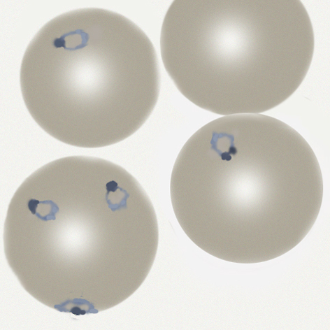
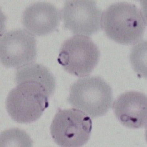
The earliest growth stage, this is characterised by fine ring forms and few other changes, this may be the only form seen in this species:
- Ring forms that are fine and delicate
- Frequently the red cells contain multiple parasites
- Parasites may have a distinctive "double dot" or signet ring form
- Parasites may appear on the accolé forms that appear flattened against the cell membrane
- Affected red cells have normal size and haemoglobin content
| The late trophozoite |
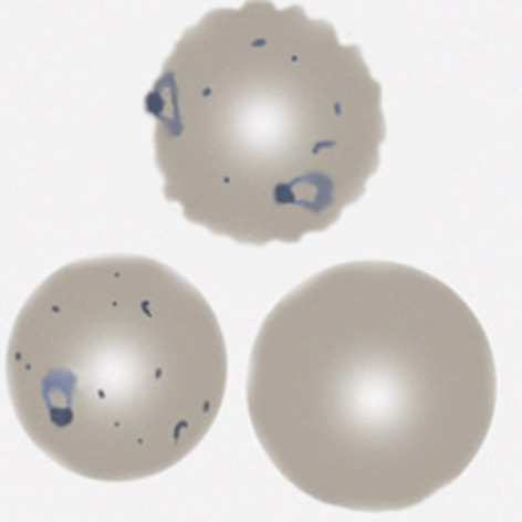
The later growth stage where parasites begin to modify the erythrocyte, causing characteristic changes with added dots and minr changes to red cell form:
- Parasites resemble early ring forms, but are thicker and may be slightly larger
- Additional blue/grey dots and clefts are seen in red cell cytoplasm when stained correctly
- These dots have low number a characteristic "dot" or "line" form Maurer's dots and clefts
- Size and shape of infected red cells is usually unaffected, but may become crenated
- The double dot, accolé, and multiple parasite forms remain present
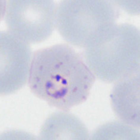
| The schizont |
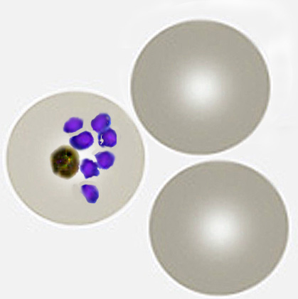
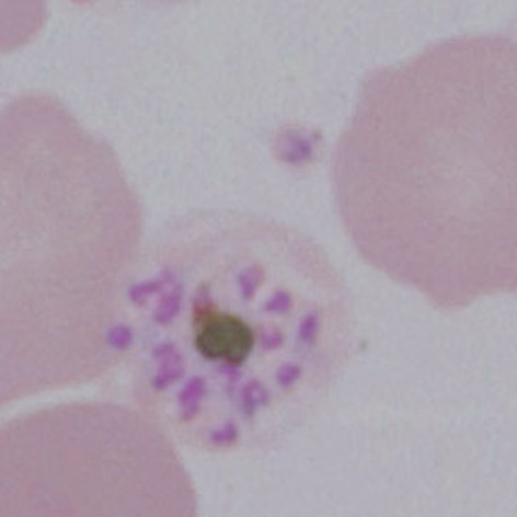
The schizont is the asexual form of the malaria parasite in blood - for a detailed description see the "Biology of malaria" section in the main menu:
- Do not generally circulate in this species unless overwhelming infection
- The merozoites cluster "untidily" as they develop
- Schizonts develop progressively to form 8-16 merozoites when mature
- In this species the loose malaria pigment may be seen in clumps between the parasites
- Red cell size is generally unaffected but red cells become pale as haemoglobin is metabolised by the parasites
The gametocyte
| The gametocyte |
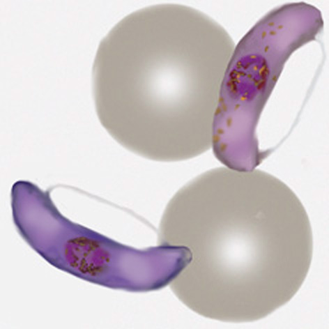
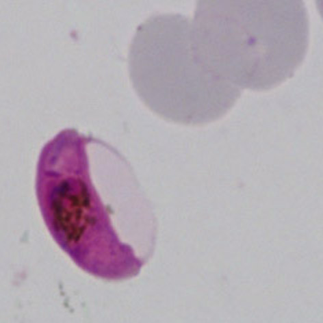
The sexual replication form (very distinctive).
- male and femaie gametocytes are elongated and have the appearance of rods
- They parasites are rod shaped but the membrane may cause them to curve into a “"banana" form”
- The residual membrane (empty of haemoglobin) is often seen as a "blister" to the side of the parasite
- The single chromatin area is in the centre of the parasite, often has pigment overlying it
- Gametocytes may not be be seen, or may be the only form present (particularly after treatment)