Maurer's dots and clefts
From haematologyetc.co.uk
Navigation
Go Back
| What are Maurer's dots and clefts?
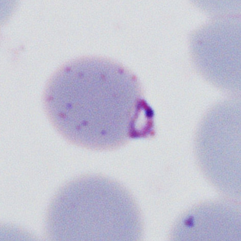
These are blue-grey dots or linear structures seen at the late trophozoites of P.falciparum. These structures lie within the erythrocyte cytoplasm and are the result of modification of the red cell by the parasite. The structures differ from the dots seen in ethrocytes infected by P.vivax or P.ovale since they are few in number (you could imagine being able to count them). They are not seen in early trophozoites so define the late trophozoite stage.
The most frequent form - two early trophozoites of P.falciparum in a single erythrocyte
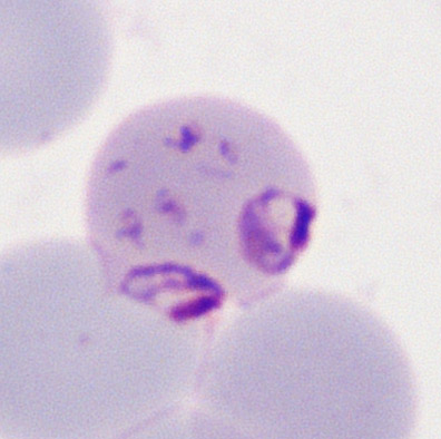
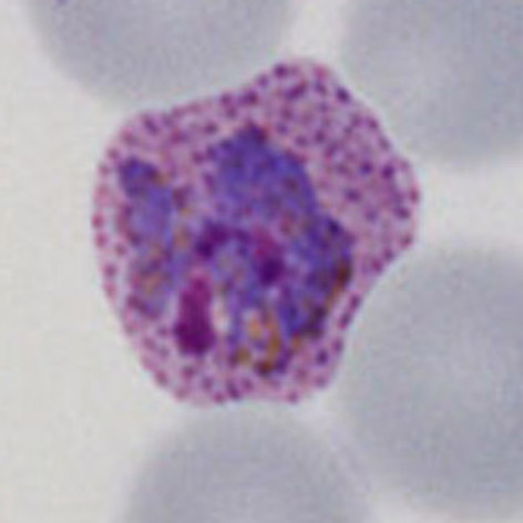
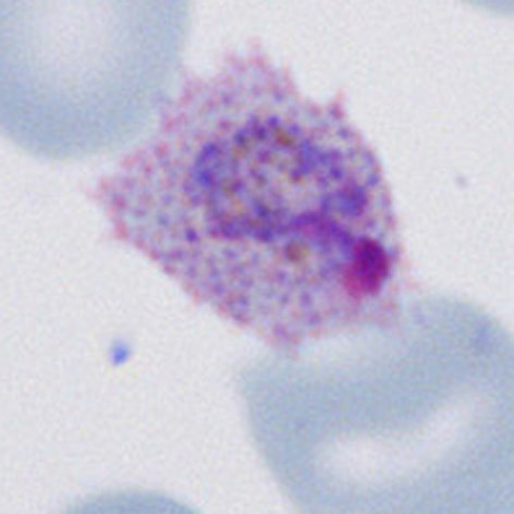
Species significance Maurer's dots and clefts are restricted to P.falciparum and can be distinguished from Schüffner's dots of P.vivax or the James' dots of P.ovale by their blue-grey colour, higher density and sometimes elongated (clefted) shape. Compare with:
Maurer's dots and clefts in a late trophozoite of P.falciparum with two tropozoites and a mixture of dots and clefts (A). These are more dense and blue grey than the more numerous, softer, amd red/purple coloured Schüffner's dots seen in P.vivax (B) and James' dots in P.ovale (C) |