Pappenheimer Bodies
From haematologyetc.co.uk
Derivation: Described by Alwin M. Pappenheimer US pathologist 1878-1955
Appearance
Grey/blue inclusions of varying size, generally small and usually (but not always) near the edge of erythrocytes. Frequently they form clusters of 2 or 3 inclusions. They are darker than Howell Jolly bodies.
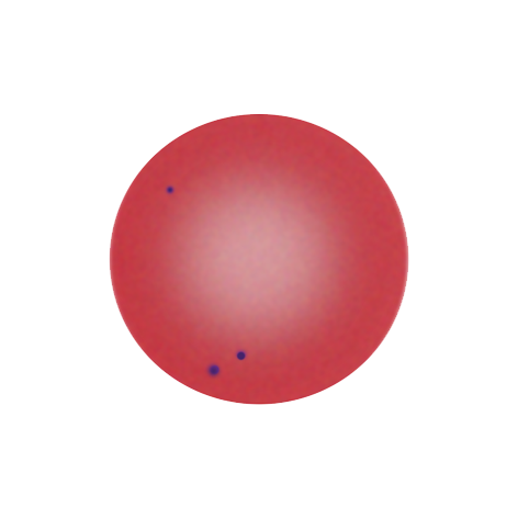
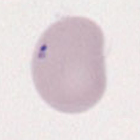
Images Small numbers (generally 1-5) small dark purple or grey inclusions, peripherally distributed and often appearing to be in groups. The red cell may be normal in size and shape or may show signs of the intrinsic cause (e.g. dysplasia, iron-loading conditions or splenectomy).
Significance
Small numbers of Pappenheimer Bodies may be seen in normal blood, particularly within polychromatic cells. When they are present in large number there will be a cause, look for hyposplenic features, or for features of pathological states that have iron-loading or aberrant iron metabolism.
Pitfalls
Pappenheimer bodies should be distinguished from other basophilic intracellular inclusions. Distinction from Howell Jolly bodies may cause particular problems, as both bodies may arise together, but generally are distinguished by attention to colour, texture and position. Platelets or stain debris overlying the erythrocyte may occasionally cause confusion, particularly the latter (see the specific section on artefacts). If there is doubt then perform an iron stain to confirm the iron content of the Pappenheimer body (see images).
Causes
| NORMAL INDIVIDUALS |
|---|
| Normal red cells may contain Pappenheimer bodies, particularly within reticulocytes. These physiological inclusions are removed by the spleen and so are not generally seen in mature erythrocytes. |
| PATHOLOGICAL (hyposplenism) |
| Following splenectomy or in hyposplenic conditions Pappenheimer bodies are not cleared from the circulating cells |
| PATHOLOGICAL (ineffective erythropoiesis) |
| Particularly where iron incorporation into haem is disturbed, or when iron loading is excessive |
| Haemoglobinopathies (thalassaemia), sideroblastic anaemia, vitamin B12 deficiency, vitamin B6 deficiency, alcohol excess, anti-pyridoxine therapy, chloramphenicol, mitochondrial disorders |
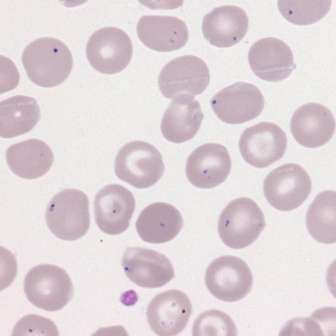
Clinical Image 1 Erythrocytes containing mainly single but in some cases multiple Pappenheimer bodies, some in groups. Note in this case the context of multiple target cells, a folded cell and excessive variation of shape. Clinical disorder: thalassaemia with iron overload.
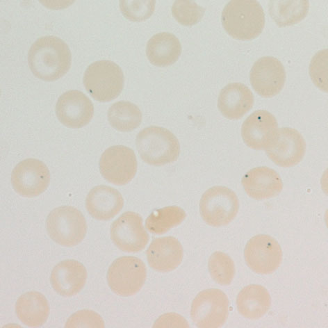
Clinical Image 2 Pearls stain for iron (same case as image 1, note the similarities of cell form). The Pappenheimer bodies stain as distinct iron-containing dots within (but not between the erythrocytes).
Pathobiology
Where erythropoiesis is stressed or ineffective (see Causes), iron may accumulate in the mitochondria of red cell precursors – this accumulated iron can be detected as “siderotic granules” using specific iron-stains. These iron-laden organelles may be retained within mature erythrocytes where they aggregate with degenerate ribosomes, lysosomes and other cell remnants. These combined aggregated proteins (not the iron itself) stain blue with Wright or Geimsa stains as Pappenheimer bodies. Like other inclusions Pappenheimer bodies are removed from red cells in the spleen, so are more prominent when the spleen is absent or dysfunctional.