Keratocytes
From haematologyetc.co.uk
Clinical Examples
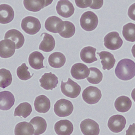
Clinical Image 1:
Typical keratocyte forms, most have two horns surrounding a central depression. There are also some echinocytes present. Clinical diagnosis: Oxidative damage and Heinz body removal
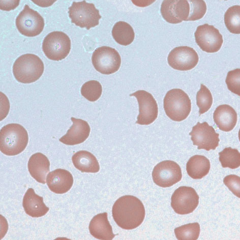
'Clinical Image 2:
Further typical keratocyte forms, note a cell with three horns (*) surrounding two central depressions. Here there is more extensive evidence of additional erythrocyte damage – (echinocytes, spherocytes and early fragments) and absent platelets. Clinical diagnosis: Thrombotic thrombocytopenic purpura
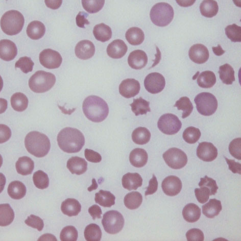
Clinical Image 3:
In this case damage is considerable, and typical keratocytes are no longer present – being replaced by fragmented cells, however same pathological origin can still be detected with residual bites and horns. Note also the polychromatic cells formed by the marrow response to cell destruction. Clinical diagnosis: Thrombotic thrombocytopenic purpura