Howell Jolly bodies
From haematologyetc.co.uk
Derivation: Eponymous: William Howell (US) and Justin Jolly (Fr) early morphologists studying effects of splenic absence
Appearance
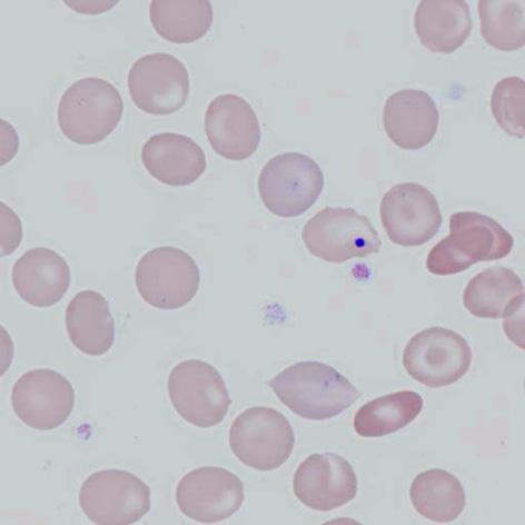
Small blue/purple fragments generally rounded – their colour and position reflects their origin as retained fragments of the cell nucleus that most often they are single and relatively centrally placed. They have relatively large size compared with other inclusions (usually 0.5-1.0μM in diameter compared with a typical erythrocyte diameter of 7.5μM).
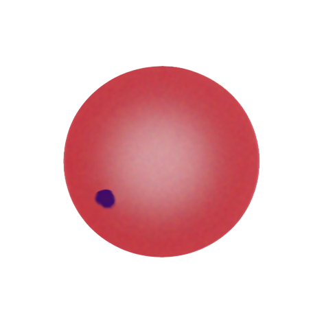
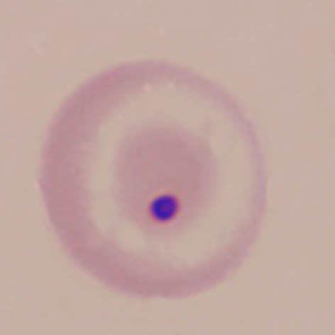
Images: In this case a single HJ body is indicated – note the relatively large size, and purple colour. This example is offset from the centre but not peripheral – a fairly typical position for these inclusions. In these examples the Howell Jolly body is accompanied by other features of hyposplenism). Look for irregularly contracted cells, target cells or speculated cells or for possible cause of hyposplenism e.g. sickle cells.
Significance
Howell-Jolly Bodies most commonly arise when spleen is absent or spleen function is impaired (hyposplenia). Occasional Howell Jolly bodies may arise in physiological conditions (see table).
Pitfalls
The group of conditions that have basophilic inclusions includes basophilic stippling and Pappenheimer bodies but these rarely cause any diagnostic confusion. Remember also however that (depending on the underlying condition) both Pappenheimer bodies and basophilic stippling may be found together with Howell Jolly bodies. More frequently, platelets or stain debris overlying red cells will cause confusion – if there is doubt look for the typical colour and rounded nature, then compare them with any platelets or debris visible between red cells.
Causes
| HYPO'SPLENISM: Physiological |
|---|
| Causes including normal i1ndividuals (very infrequent), pregnancy (infrequent), neonates (frequent) – more common in hyposplenic states |
| HYPOSPLENISM: Pathological absence |
| Congenital asplenism, |
| HYPOSPLENISM: Pathological hypofunction |
| Including: Normal neonates, Coeliac Disease, Bone marrow transplant, Pregnancy |
Clinical Examples
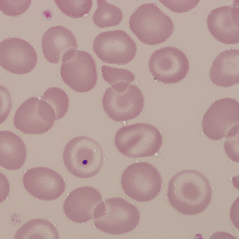
Clinical Image 1: One almost central Howell Jolly body that is almost centrally placed – recognise by the size and colour. In this film there are also very frequent target cells and contracted cells. Clinical condition: HbSC disease with hyposplenic features
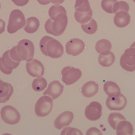
Clinical Image 2: Very frequent Howell Jolly bodies in cells showing a range of other abnormal features (excessive variation in shape and size). Note the range of sizes and positions. Clinical condition: Post-splenectomy
Clinical Image 3: A single cell containing a Howell Jolly body, in this case there are accompanying target cells, one “boat shaped” and several dense contracted cells. Clinical condition: sickle cell disease with hyposplenism (no true sickle cells are shown on this film).
Pathobiology
When red cell formation is completed the nucleus is extruded. Before this the nuclear material becomes condensed (pyknosis) and often fragmented (karryohexis) leaving many separate remnants. Normally these nuclear remains are efficiently removed from circulating erythrocytes by the spleen, however if splenic function is impaired the fragments remain as Howell Jolly bodies.