Flow cytometry: Chronic Lymphocytic leukaemia: Difference between revisions
From haematologyetc.co.uk
No edit summary |
No edit summary |
||
| Line 3: | Line 3: | ||
[[file:CLL.jpg|200px|link=CLL.jpg]] | [[file:CLL.jpg|200px|link=CLL.jpg]] | ||
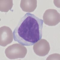
<span style="size: 90%>''Typical CLL morphology, note the small/medium size, bland chromatin pattern in a slightly squared nucleus, and the tendency of cytoplasm to gently engage adjacent red cells''</span> | <span style="font-size:90%">''Typical CLL morphology, note the small/medium size, bland chromatin pattern in a slightly squared nucleus, and the tendency of cytoplasm to gently engage adjacent red cells''</span> | ||
| Line 21: | Line 21: | ||
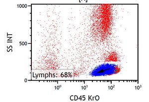
<span style="size: 90%>''Side scatter of CLL cells is typically low, with a broad range and relatively weak expression of CD45 compared with normal lymphocytes. The CLL cells (highlighted in blue) therefore tend to the left of normal T cells (red) - giving the CLL population a rather elongated "slug-like" shape.'' | <span style="font-size:90%">''Side scatter of CLL cells is typically low, with a broad range and relatively weak expression of CD45 compared with normal lymphocytes. The CLL cells (highlighted in blue) therefore tend to the left of normal T cells (red) - giving the CLL population a rather elongated "slug-like" shape.'' | ||
Revision as of 15:05, 28 June 2023
Typical CLL morphology, note the small/medium size, bland chromatin pattern in a slightly squared nucleus, and the tendency of cytoplasm to gently engage adjacent red cells
Background to flow cytometric diagnosis
Typical CLL can be diagnosed with some confidence using flow cytometry.
However, individual markers may not be expressed in all cases - this can cause diagnostic difficulty particularly in distinguishing CLL from atypical cases of MCL or MZL. If this is the case then the atypical features should be reported, and the possibility of alternative diagnoses acknowledged.
Note also that cases with a low lymphocytosis (<1x10⁹) may represent monoclonal B-lymphocytosis with CLL phenotype (click here for more).
CD45 Expression
Side scatter of CLL cells is typically low, with a broad range and relatively weak expression of CD45 compared with normal lymphocytes. The CLL cells (highlighted in blue) therefore tend to the left of normal T cells (red) - giving the CLL population a rather elongated "slug-like" shape.
| Immunophenotype of CLL | |||
|---|---|---|---|
| Major markers useful in CLL diagnosis | |||
| Marker | Freq | Level | Comment |
| CD19 | - | Expression is expected, but less strong than normal B cells | |
| κ/λ | Expect weak restricted κ or λ or absent expression | ||
| CD5 | - | Characteristic of CLL, usually less strong than on T cells | |
| CD23 | - | Characteristic of CLL, although can rarely be weak or absent | |
| CD79b | wk | Expression is expected, but characteristically weak | |
| CD200 | - | Characteristically positive in CLL and negative in MCL | |
| FMC7 | May be expressed by atypical cases, generally absent | ||
| Other relevant markers | |||
| CD10 | - | Infrequent in CLL, consider FL if detected | |
| CD11C | wk | Expressed in some cases, but tends to be weak | |
| CD20 | wk | Expression is expected, but characteristically weak | |
| CD25 | wk | Expressed by many cases of CLL but characteristically weak | |
| CD38 | wk | Expressed by some cases of CLL, typically weak | |
| CD43 | - | Similar to CD200, may be helpful in some cases | |
| CD103 | - | Not expressed by CLL, consider HCL if found | |
| CD138 | - | Predominantly expressed at plasma cell differentiation | |
Key to table:
Markers most useful in initial diagnosis are underlined in blue text
Key to colour code for expression frequency Click for link
Key to expression strength code and use Click for link

