Banana gametocyte: Difference between revisions
From haematologyetc.co.uk
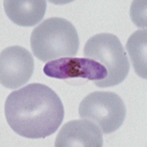
(Created page with " ---- <gallery mode="nolines" widths="220px" heights="220px" > File:MB1.jpg|: ''P.falciparum'' macrogametocyte|link={{filepath:MB1.jpg}} File:MB2.jpg|: ''P.falciparum microgametocyte''|link={{filepath:MB2.jpg}} </gallery> <span style="color:navy>'''Description'''</span> The gametocyte stage of ''P.falciparum'' has the form of a round-ended rod. The rod is slightly longer than a red cell so becomes curved by the red cell membrane and is often described a "banana shap...") |
No edit summary |
||
| (2 intermediate revisions by the same user not shown) | |||
| Line 1: | Line 1: | ||
---- | |||
'''Navigation'''</br> | |||
[[Plasmodium falciparum: Morphology|Go Back]] | |||
---- | |||
--- | {| class="wikitable" style="border-style: solid; border-width: 4px; color:black" | ||
|colspan="1" style = "font-size:100%; color:black; background: FFFAFA"|<span style="color:navy>'''What a "banana" gametocyte?'''</span> | |||
<gallery mode="nolines" widths="220px" heights="220px" > | <gallery mode="nolines" widths="220px" heights="220px" > | ||
File:MB1.jpg|: ''P.falciparum'' macrogametocyte|link={{filepath:MB1.jpg}} | File:MB1.jpg|: ''P.falciparum'' macrogametocyte|link={{filepath:MB1.jpg}} | ||
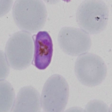
File: | File:MBi2.jpg|: ''P.falciparum microgametocyte''|link={{filepath:MBi2.jpg}} | ||
</gallery> | </gallery> | ||
| Line 31: | Line 36: | ||
This is a squashed and distorted trophozoite of ''P.vivax'' that has been compressed by the red cells around it. The | This is a squashed and distorted trophozoite of ''P.vivax'' that has been compressed by the red cells around it. The signs of external compression and but also the presence of Schüffner's dots in the cytoplasm of the red cell should make it clear that this is not ''P.falciparum''. | ||
Latest revision as of 12:45, 30 March 2024
Navigation
Go Back
What a "banana" gametocyte?
Additional images
|