''P.falciparum'' gallery: Difference between revisions
From haematologyetc.co.uk
No edit summary |
No edit summary |
||
| (11 intermediate revisions by the same user not shown) | |||
| Line 11: | Line 11: | ||
<span style="font-size:95%">At this stage we look for typical (and often frequent) delicate rings within red cells that have normal (or slightly crenated) appearance. Forms often seen in this species include accolé forms, double chromatin dot forms, and multiple parasites within infected red cells. | <span style="font-size:95%">At this stage we look for typical (and often frequent) delicate rings within red cells that have normal (or slightly crenated) appearance. Forms often seen in this species include accolé forms, double chromatin dot forms, and multiple parasites within infected red cells. | ||
<gallery mode="traditional" widths=240px heights=240px> | <gallery mode="traditional" widths=240px heights=240px> | ||
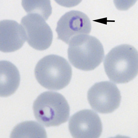
File: | File:PFET1p.jpg|<span style="font-size:80%">'''Fine ring form''' The small and delicate form of this species</span>|link={{filepath:PFET1p.jpg}} | ||
File: | File:PFET2p.jpg|<span style="font-size:80%">'''Double chromatin dot form''' Two chromatin dots (sometimes known as "signet ring" form).</span>|link={{filepath:PFET2p.jpg}} | ||
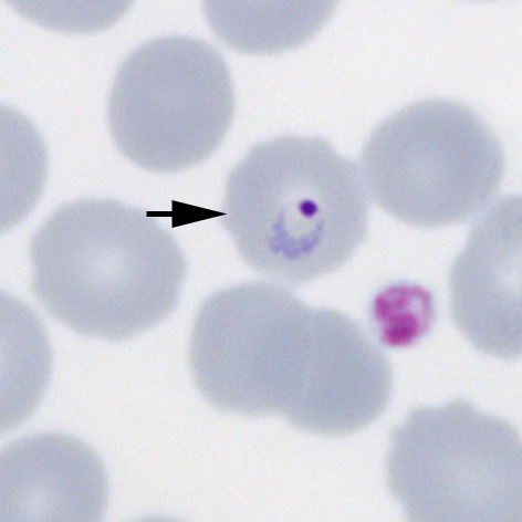
File: | File:PFET3p.jpg|<span style="font-size:80%">'''Accolé form''': The arrowed form is closely associated with the red cell membrane</span>|link={{filepath:PFET3p.jpg}} | ||
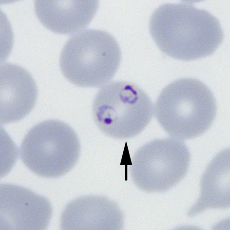
File: | File:PFET4p.jpg|<span style="font-size:80%">'''Multiple parasites''' Two parasites within a single red cells (arrowed)</span>|link={{filepath:PFET4p.jpg}} | ||
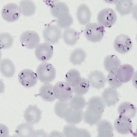
File: | File:PFET5p.jpg|<span style="font-size:80%">'''High parasitaemia''' Most of the typical ''P.falciparum'' forms are present</span>|link={{filepath:PFET5p.jpg}} | ||
</gallery>" | |||
</gallery> | |||
---- | ---- | ||
Latest revision as of 15:35, 5 March 2024
P.falciparum early trophozoites
Summary
At this stage we look for typical (and often frequent) delicate rings within red cells that have normal (or slightly crenated) appearance. Forms often seen in this species include accolé forms, double chromatin dot forms, and multiple parasites within infected red cells.

Fine ring form The small and delicate form of this species
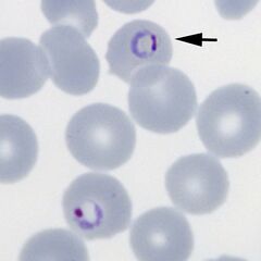
Double chromatin dot form Two chromatin dots (sometimes known as "signet ring" form).
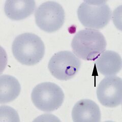
Accolé form: The arrowed form is closely associated with the red cell membrane
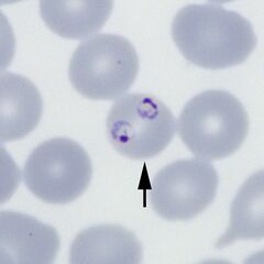
Multiple parasites Two parasites within a single red cells (arrowed)
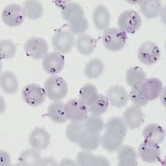
High parasitaemia Most of the typical P.falciparum forms are present

"