''P.falciparum'' gallery: Difference between revisions
From haematologyetc.co.uk
No edit summary |
No edit summary |
||
| Line 13: | Line 13: | ||
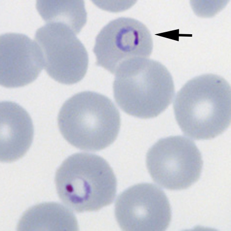
File:PFET1p.jpg|<span style="font-size:80%">'''Fine ring form''' The small and delicate form of this species</span>|link={{filepath:PFET2p.jpg}} | File:PFET1p.jpg|<span style="font-size:80%">'''Fine ring form''' The small and delicate form of this species</span>|link={{filepath:PFET2p.jpg}} | ||
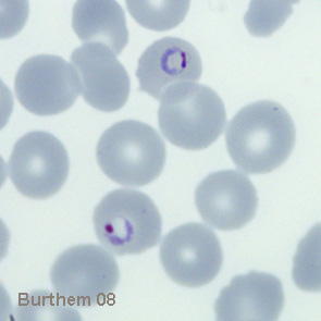
File:PFET2p.jpg|<span style="font-size:80%">'''Double chromatin dot form''' Two chromatin dots (sometimes known as "signet ring" form).</span>|link={{filepath:PFETi2.jpg}} | File:PFET2p.jpg|<span style="font-size:80%">'''Double chromatin dot form''' Two chromatin dots (sometimes known as "signet ring" form).</span>|link={{filepath:PFETi2.jpg}} | ||
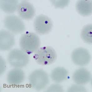
File:PFET3p.jpg|<span style="font-size:80%">Multiple parasites in a single erythrocyte (one double chromatin dot form)</span>|link={{filepath:PFET3p.jpg}} | File:PFET3p.jpg|<span style="font-size:80%">'''Multiple parasites''': two in a single erythrocyte (one double chromatin dot form)</span>|link={{filepath:PFET3p.jpg}} | ||
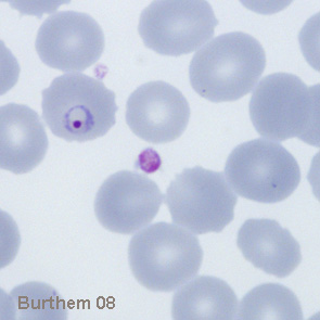
File:PFETi4.jpg|<span style="font-size:80%">Delicate and small early trophozoite form of ''Plasmodium falciparum''</span>|link={{filepath:PFETi4.jpg}} | File:PFETi4.jpg|<span style="font-size:80%">Delicate and small early trophozoite form of ''Plasmodium falciparum''</span>|link={{filepath:PFETi4.jpg}} | ||
File:PFETi5.jpg|<span style="font-size:80%">Two early forms of ''P.falciparum'' one with the characteristic double chromatin dot</span>|link={{filepath:PFETi5.jpg}} | File:PFETi5.jpg|<span style="font-size:80%">Two early forms of ''P.falciparum'' one with the characteristic double chromatin dot</span>|link={{filepath:PFETi5.jpg}} | ||
Revision as of 13:56, 2 March 2024
P.falciparum early trophozoites
Summary
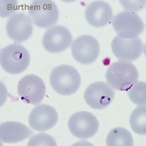
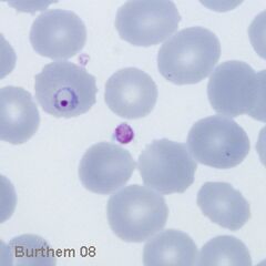
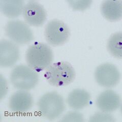
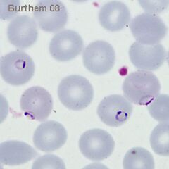
At this stage we look for typical (and often frequent) delicate rings within red cells that have normal (or slightly crenated) appearance. Forms often seen in this species include accolé forms, double chromatin dot forms, and multiple parasites within infected red cells.
Fine ring form The small and delicate form of this species
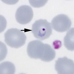
Double chromatin dot form Two chromatin dots (sometimes known as "signet ring" form).
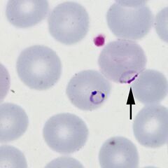
Multiple parasites: two in a single erythrocyte (one double chromatin dot form)
Delicate and small early trophozoite form of Plasmodium falciparum
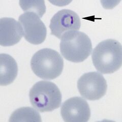
Two early forms of P.falciparum one with the characteristic double chromatin dot
Multiple parasites in a single erythrocyte (one double chromatin dot form)