Gallery of early trophozoites: Difference between revisions
From haematologyetc.co.uk
No edit summary |
No edit summary |
||
| (68 intermediate revisions by the same user not shown) | |||
| Line 1: | Line 1: | ||
---- | ---- | ||
'''Navigation'''</br> | '''Navigation'''</br> | ||
[[ | [[Galleries|Go Back]] | ||
---- | ---- | ||
<span style="font-size:95% | <span style="font-size:95%">'''General Comments:''' At the very earliest point all trophozoites appear as ring forms and species differences are very difficult to distinguish - some "species specific" features may appear as parasites mature toward late trophozoite stages.</br></br> | ||
<span style="font-size: | |||
---- | |||
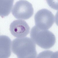
<span style="font-size:90%">''' ''P.falciparum'' '''</span></br> | |||
<span style="font-size:90%">Small delicate rings, and these '''may be the only forms seen in some patients at diagnosis'''. Infected red cells have normal (or slightly crenated) appearance.</br> | |||
<gallery | <gallery heights=200px widths=200px> | ||
File:PFET1g.jpg|<span style="font-size:80%">Fine ring form</span>|link={{filepath:PFET1g.jpg}} | File:PFET1g.jpg|<span style="font-size:80%">Fine ring form</span>|link={{filepath:PFET1g.jpg}} | ||
File:PFET2g.jpg|<span style="font-size:80%">Double dot form and normal ring</span>|link={{filepath:PFET2g.jpg}} | File:PFET2g.jpg|<span style="font-size:80%">Double dot form and normal ring</span>|link={{filepath:PFET2g.jpg}} | ||
| Line 13: | Line 16: | ||
File:PFET4g.jpg|<span style="font-size:80%">Multiple parasite form</span>|link={{filepath:PFET4g.jpg}} | File:PFET4g.jpg|<span style="font-size:80%">Multiple parasite form</span>|link={{filepath:PFET4g.jpg}} | ||
</gallery>" | </gallery>" | ||
---- | |||
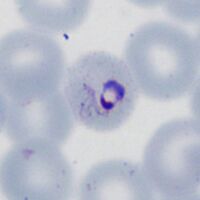
<span style="font-size:95%">''' ''P.vivax'' '''</span></br> | |||
<span style="font-size:90%">Rings begin as small forms, but become larger asociated with enlarged distorted red cells as they develop. Schüffner's dots will become present | |||
<gallery heights=200px widths=200px> | |||
File:PVET1g.jpg|<span style="font-size:80%">Early ring form</span>|link={{filepath:PVET1g.jpg}} | |||
File:PVET2g.jpg|<span style="font-size:80%">Early ring form with faint dots</span>|link={{filepath:PVET2g.jpg}} | |||
File:PVET3g.jpg|<span style="font-size:80%">Llarge thickened ring trophozoite</span>|link={{filepath:PVET3g.jpg}} | |||
File:PVET4g.jpg|<span style="font-size:80%">Distorted ting trophozoite and red cell, marked Schüffner's dots</span>|link={{filepath:PVET4g.jpg}} | |||
</gallery>" | |||
---- | |||
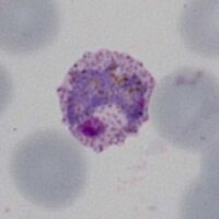
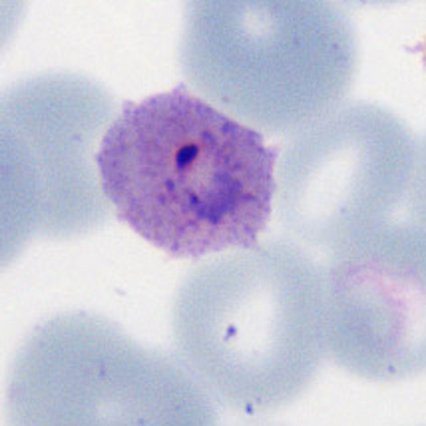
<span style="font-size:90%">''' ''P.ovale'' '''</span></br> | |||
<span style="font-size:90%">Ring form is retained but enlarges, red cells may develop fimbriation and enlarged ovoid form with visible James' dots. | |||
<gallery mode="traditional" widths=200px heights=200px> | |||
File:POET1g.jpg|<span style="font-size:80%">Early ring form</span>|link={{filepath:POET1g.jpg}} | |||
File:POET2g.jpg|<span style="font-size:80%">Ring with dots/fimbriation</span>|link={{filepath:POET2g.jpg}} | |||
File:POET3g.jpg|<span style="font-size:80%">faint Ziemann's dots</span>|link={{filepath:POET3g.jpg}} | |||
File:POET4g.jpg|<span style="font-size:80%">Ring early ovoid change</span>|link={{filepath:POET4g.jpg}} | |||
</gallery>" | |||
---- | |||
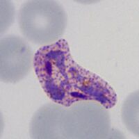
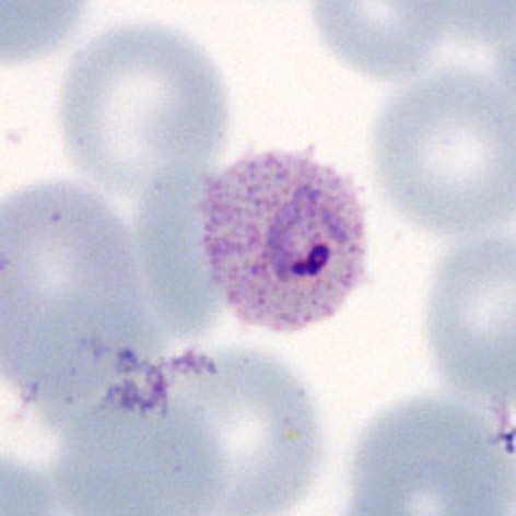
<span style="font-size:90%">''' ''P.malariae'' '''</span></br> | |||
<span style="font-size:90%">Infected red cells are generally infrequent. Early trophozoites are small in normal or small erythrocytes, and may have central chromatin dot, elongation or angular forms. | |||
<gallery mode="traditional" widths=200px heights=200px> | |||
File:MET1g.jpg|<span style="font-size:80%">Ring form in small red cell</span>|link={{filepath:MET1g.jpg}} | |||
File:MET2g.jpg|<span style="font-size:80%">The central chromatin dot</span>|link={{filepath:MET2g.jpg}} | |||
File:PMET3g.jpg|<span style="font-size:80%">Early elongation, Stinton's dots</span>|link={{filepath:MET3g.jpg}} | |||
File:MET4g.jpg|<span style="font-size:80%">Early angularity of form</span>|link={{filepath:MET4g.jpg}} | |||
</gallery>" | |||
---- | |||
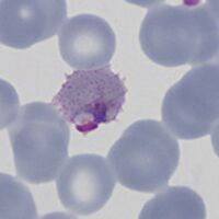
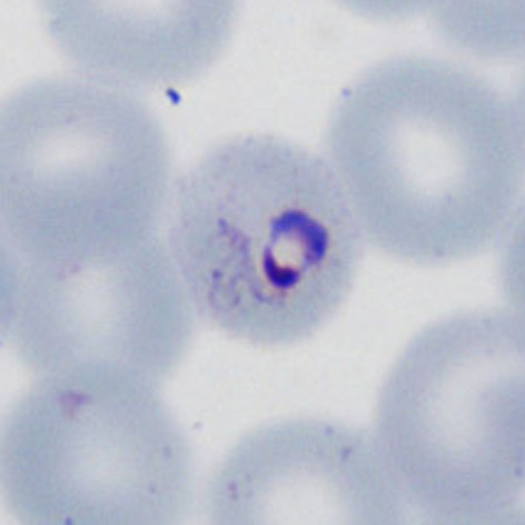
<span style="font-size:90%">''' ''P.knowlesi'' '''</span></br> | |||
<span style="font-size:90%">The early trophozoite may resembles ''P.falciparum'' and infected cells may be frequent. Later forms however begin to resemble parasites of ''P.malariae''. | |||
<gallery mode="traditional" widths=200px heights=200px> | |||
File:PKET1.jpg|<span style="font-size:80%">Fine early rings</span>|link={{filepath:PKET1.jpg}} | |||
File:PKET2a.jpg|<span style="font-size:80%">Double dot (right)</span>|link={{filepath:PKET2a.jpg}} | |||
File:PKET3a.jpg|<span style="font-size:80%">Accolé form</span>|link={{filepath:PKET3a.jpg}} | |||
File:PKET4a.jpg|<span style="font-size:80%">Multiple infection</span>|link={{filepath:PKET4a.jpg}} | |||
</gallery> | |||
---- | ---- | ||
Latest revision as of 13:30, 27 November 2024
Navigation
Go Back
General Comments: At the very earliest point all trophozoites appear as ring forms and species differences are very difficult to distinguish - some "species specific" features may appear as parasites mature toward late trophozoite stages.
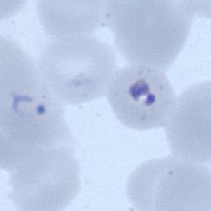
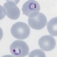
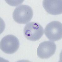
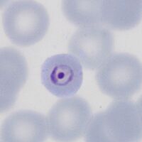
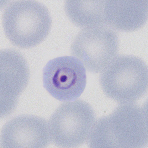
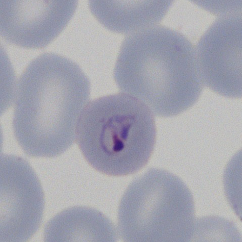
P.falciparum
Small delicate rings, and these may be the only forms seen in some patients at diagnosis. Infected red cells have normal (or slightly crenated) appearance.
Fine ring form
Double dot form and normal ring

Accolé and double dot forms
Multiple parasite form


"
P.vivax
Rings begin as small forms, but become larger asociated with enlarged distorted red cells as they develop. Schüffner's dots will become present
Early ring form
Early ring form with faint dots
Llarge thickened ring trophozoite
Distorted ting trophozoite and red cell, marked Schüffner's dots


"
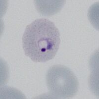
P.ovale
Ring form is retained but enlarges, red cells may develop fimbriation and enlarged ovoid form with visible James' dots.
Early ring form

Ring with dots/fimbriation
faint Ziemann's dots

Ring early ovoid change

"
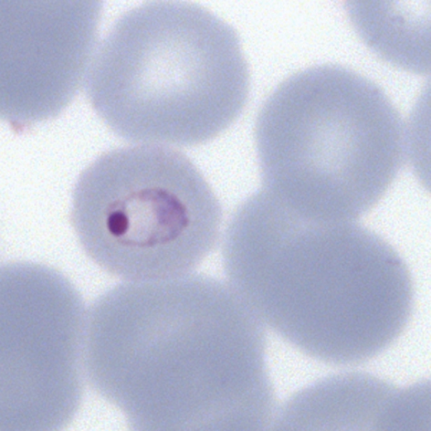
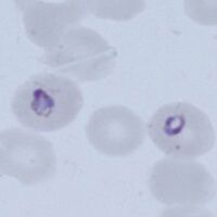
P.malariae
Infected red cells are generally infrequent. Early trophozoites are small in normal or small erythrocytes, and may have central chromatin dot, elongation or angular forms.
Ring form in small red cell
The central chromatin dot

Early elongation, Stinton's dots
Early angularity of form
"
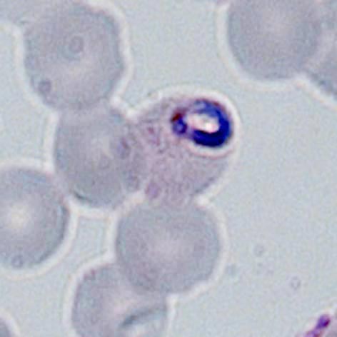
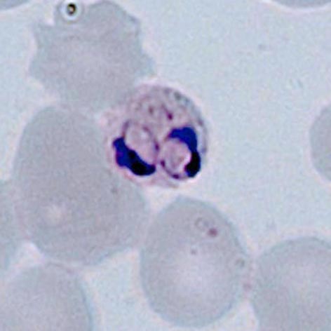
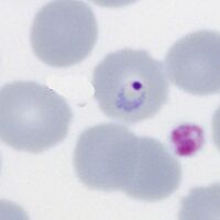
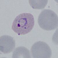
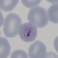
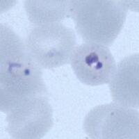
P.knowlesi
The early trophozoite may resembles P.falciparum and infected cells may be frequent. Later forms however begin to resemble parasites of P.malariae.
Fine early rings
Double dot (right)

Accolé form

Multiple infection