Species identification: summary page: Difference between revisions
From haematologyetc.co.uk
(Created page with "Return to Malaria Index Page'' ---- {| class="wikitable" style="border-style: solid; border-width: 5px; border-color: #bcd4e6; color:black" |colspan="1" style = "font-size:90%; color:black; background: #bcd4e6"|'''''Plasmodium falciparum''''' |} <span style="font-size:95%">'''Summary'''</span> *<span style="font-size:95%">Small and fine ring forms, may have accolé (edge) forms or multiple parasites per cell</span> *<span style="font-size:95%">Mau...") |
No edit summary |
||
| (100 intermediate revisions by 2 users not shown) | |||
| Line 1: | Line 1: | ||
[[Malaria Index| | ---- | ||
'''Navigation'''</br> | |||
<span style="font-size:80%">(click blue highlighted text to return to page)</span></br></br> | |||
<span style="font-size:90%">[[Malaria Index|Malaria main index]]''</span></br> | |||
<span style="font-size:90%">>This page: <u>Species Indentification: summary</u></span> | |||
---- | ---- | ||
{| class="wikitable" style="border-style: solid; border-width: 5px; border-color: #023020; color:black" | |||
{| class="wikitable" style="border-style: solid; border-width: 5px; border-color: # | |colspan="1" style = "font-size:100%; color:black; background: #afbddb |'''''Plasmodium falciparum''''' | ||
|colspan="1" style = "font-size: | |||
|} | |} | ||
<gallery mode="nolines" widths=140px heights=150px> | |||
<gallery mode="nolines" widths= | |||
File:PFETc.jpg|<span style="font-size:80%">''Early trophozoite''</span>|link={{filepath:PFETc.jpg}} | File:PFETc.jpg|<span style="font-size:80%">''Early trophozoite''</span>|link={{filepath:PFETc.jpg}} | ||
File:PFLTc.jpg|<span style="font-size:80%">Late trophozoite</span>|link={{filepath:PFLTc.jpg}} | File:PFLTc.jpg|<span style="font-size:80%">Late trophozoite</span>|link={{filepath:PFLTc.jpg}} | ||
File: | File:PFSc2.jpg|<span style="font-size:80%">Schizont (rare)</span>|link={{filepath:PFSc2.jpg}} | ||
File:PFGc.jpg|<span style="font-size:80%">Gametocyte</span>|link={{filepath:PFGc.jpg}} | File:PFGc.jpg|<span style="font-size:80%">Gametocyte</span>|link={{filepath:PFGc.jpg}} | ||
</gallery> | </gallery> | ||
<span style="font-size:95%">'''Summary'''</span> | |||
*<span style="font-size:95%">Small and fine ring forms, look for typical forms accolé, multiple parasites per cell, double dot</span> | |||
*<span style="font-size:95%">Characteristic Maurer's dots and clefts in late trophozoites</span> | |||
*<span style="font-size:95%">The irregular and "tatty" schizonts very '''rarely seen''' in blood unless severe infection</span> | |||
*<span style="font-size:95%">Characteristic elongated (often curved) 'banana' gametocytes</span> | |||
</span> | |||
<div style="width: | |||
{| class="wikitable" style="border-left:solid 4px | <div style="width: 230px"> | ||
| colspan="1"''|[[Plasmodium falciparum: Morphology| | {| class="wikitable" style="border-left:solid 4px gray;border-right:solid 4px gray;border-top:solid 4px gray;border-bottom:solid 4px gray; font-size:90%; color:navy; align:center" | ||
| colspan="1"''|[[Plasmodium falciparum: Morphology|'''CLICK''' for detailed description]]'' | |||
|} | |} | ||
</div> | </div> | ||
| Line 36: | Line 37: | ||
<div style="width: 95%"> | <div style="width: 95%"> | ||
{| class="wikitable" style="border-style: solid; border-width: 5px; color:black" | {| class="wikitable" style="border-style: solid; border-width: 5px; border-color: #023020; color:black" | ||
|colspan="1" style = "font-size: | |colspan="1" style = "font-size:100%; color:black; background: #afbddb |'''''Plasmodium vivax''''' | ||
|} | |} | ||
<gallery mode="nolines" widths=140px heights=140px> | |||
File:PVETc.jpg|<span style="font-size:80%">''Early trophozoite''</span>|link={{filepath:PVETc.jpg}} | |||
File:PVLTc.jpg|<span style="font-size:80%">Late trophozoite</span>|link={{filepath:PVLTc.jpg}} | |||
File:PVSc.jpg|<span style="font-size:80%">Schizont (rare)</span>|link={{filepath:PVSc.jpg}} | |||
File:PVGc.jpg|<span style="font-size:80%">Gametocyte</span>|link={{filepath:PVGc.jpg}} | |||
</gallery> | |||
''' | '''Summary''' | ||
*Large and robust rings that become amoeboid during later development | *Large and robust rings that become "amoeboid" during later development | ||
*Red cells become increasingly enlarged and distorted as parasites mature | *Red cells become increasingly enlarged and distorted as parasites mature | ||
*Schüffner's dots are visible in appropriately stained thin blood films | *Schüffner's dots are visible in appropriately stained thin blood films | ||
| Line 57: | Line 57: | ||
'' | <div style="width: 230px"> | ||
{| class="wikitable" style="border-left:solid 4px gray;border-right:solid 4px gray;border-top:solid 4px gray;border-bottom:solid 4px gray; font-size:90%; color:navy; align:center" | |||
| colspan="1"''|[[Plasmodium vivax: Morphology|'''CLICK''' for detailed description]]'' | |||
|} | |||
</div> | |||
---- | ---- | ||
<div style="width: 95%"> | <div style="width: 95%"> | ||
{| class="wikitable" style="border-style: solid; border-width: 5px; color:black" | {| class="wikitable" style="border-style: solid; border-width: 5px; border-color: #023020; color:black" | ||
|colspan="1" style = "font-size: | |colspan="1" style = "font-size:100%; color:black; background: #afbddb |'''''Plasmodium ovale''''' | ||
|} | |} | ||
[[File:POETc.jpg|150px|link={{filepath:POETc.jpg}}]] | [[File:POETc.jpg|150px|link={{filepath:POETc.jpg}}]] | ||
[[File:POLTc.jpg|150px|link={{filepath:POLTc.jpg}}]] | [[File:POLTc.jpg|150px|link={{filepath:POLTc.jpg}}]] | ||
| Line 76: | Line 82: | ||
'''Brief summary''' | '''Brief summary''' | ||
* | *Ring forms are large and robust and often retained in the late trophozoite stage | ||
*Red cells | *Red cells become moderately enlarged and may have oval shape with characteristic fimbriation | ||
*Schüffner's (James) dots seen in appropriately stained samples | *Schüffner's (James) dots form dusing development and will be seen in appropriately stained samples | ||
*All forms tend to circulate | *All developmental forms tend to circulate and may be difficult to distinguish from ''P.vivax'' | ||
'' | <div style="width: 230px"> | ||
{| class="wikitable" style="border-left:solid 4px gray;border-right:solid 4px gray;border-top:solid 4px gray;border-bottom:solid 4px gray; font-size:90%; color:navy; align:center" | |||
| colspan="1"''|[[Plasmodium ovale: Morphology|'''CLICK''' for detailed description]]'' | |||
|} | |||
</div> | |||
---- | ---- | ||
<div style="width: 95%"> | <div style="width: 95%"> | ||
{| class="wikitable" style="border-style: solid; border-width: 5px; color:black" | {| class="wikitable" style="border-style: solid; border-width: 5px; border-color: #023020; color:black" | ||
|colspan="1" style = "font-size: | |colspan="1" style = "font-size:100%; color:black; background: #afbddb |'''''Plasmodium malariae''''' | ||
|} | |} | ||
[[File:PMETc.jpg|150px|link={{filepath:PMETc.jpg}}]] | [[File:PMETc.jpg|150px|link={{filepath:PMETc.jpg}}]] | ||
[[File:PMLTc.jpg|150px|link={{filepath:PMLTc.jpg}}]] | [[File:PMLTc.jpg|150px|link={{filepath:PMLTc.jpg}}]] | ||
| Line 113: | Line 123: | ||
---- | ---- | ||
{| class="wikitable" style="border-style: solid; border-width: 5px; color:black" | {| class="wikitable" style="border-style: solid; border-width: 5px; border-color: #023020; color:black" | ||
|colspan="1" style = "font-size: | |colspan="1" style = "font-size:100%; color:black; background: #fff5ee |'''''Plasmodium knowlesi''''' | ||
|} | |} | ||
<gallery mode="nolines" widths=150px heights=150px> | <gallery mode="nolines" widths=150px heights=150px> | ||
File:PKETc.jpg|<span style="font-size:80%">''Early trophozoite''</span>|link={{filepath:PKETc.jpg}} | File:PKETc.jpg|<span style="font-size:80%">''Early trophozoite''</span>|link={{filepath:PKETc.jpg}} | ||
| Line 137: | Line 148: | ||
''For more information'' | ''For more information'' | ||
*[[''Plasmodium knowlesi'': Morphology|click for full description of ''P.knowlesis'' morphology]] | *[[''Plasmodium knowlesi'': Morphology|click for full description of ''P.knowlesis'' morphology]] | ||
*[[''Plasmodium knowlesi'': Morfología|haga clic para obtener una descripción completa de la morfología de ''P.knowlesis'']] | |||
*[[''Plasmodium knowlesi'': Gallery|click to visit the gallery of ''P.knowlesi'' forms]] | *[[''Plasmodium knowlesi'': Gallery|click to visit the gallery of ''P.knowlesi'' forms]] | ||
---- | ---- | ||
Latest revision as of 14:26, 1 May 2024
Navigation
(click blue highlighted text to return to page)
Malaria main index
>This page: Species Indentification: summary
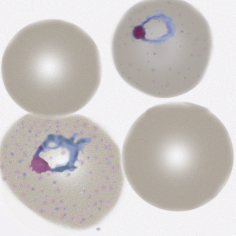
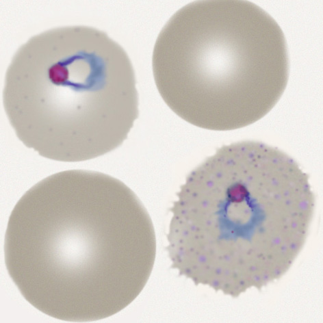
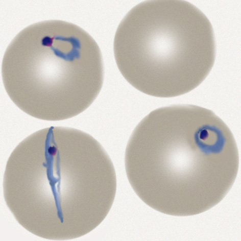
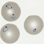
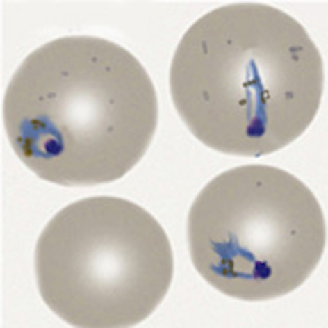
| Plasmodium falciparum |
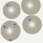
Early trophozoite
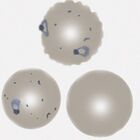
Late trophozoite

Schizont (rare)
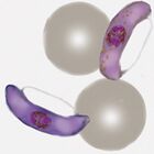
Gametocyte
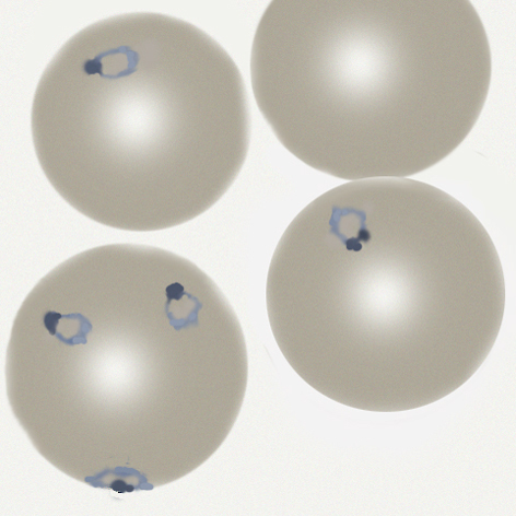

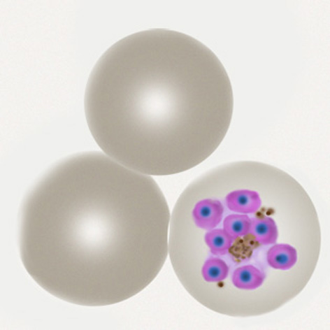

Summary
- Small and fine ring forms, look for typical forms accolé, multiple parasites per cell, double dot
- Characteristic Maurer's dots and clefts in late trophozoites
- The irregular and "tatty" schizonts very rarely seen in blood unless severe infection
- Characteristic elongated (often curved) 'banana' gametocytes
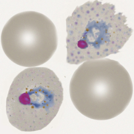
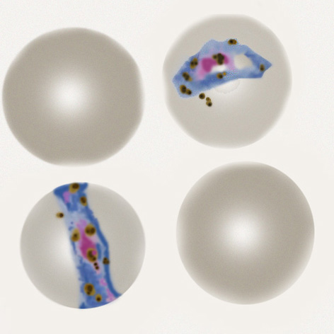
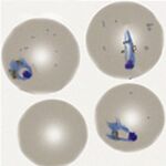
| Plasmodium vivax |
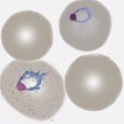
Early trophozoite
Late trophozoite

Schizont (rare)
Gametocyte
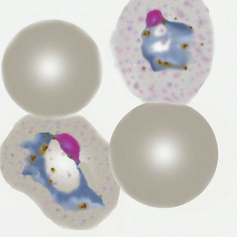
Summary
- Large and robust rings that become "amoeboid" during later development
- Red cells become increasingly enlarged and distorted as parasites mature
- Schüffner's dots are visible in appropriately stained thin blood films
- All forms tend to circulate with large schizont and gametocyte forms present
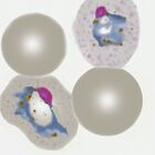
| Plasmodium ovale |
Brief summary
- Ring forms are large and robust and often retained in the late trophozoite stage
- Red cells become moderately enlarged and may have oval shape with characteristic fimbriation
- Schüffner's (James) dots form dusing development and will be seen in appropriately stained samples
- All developmental forms tend to circulate and may be difficult to distinguish from P.vivax
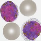
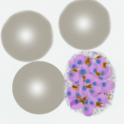
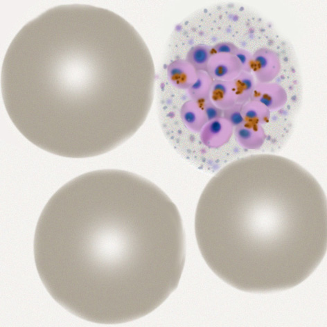
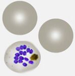
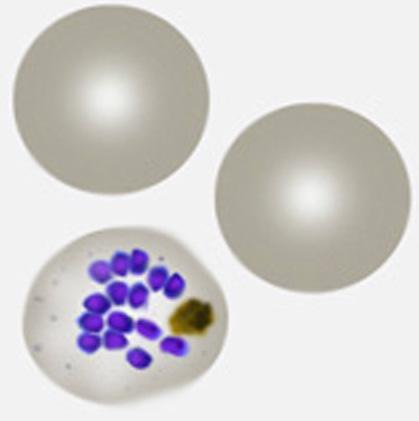
| Plasmodium malariae |
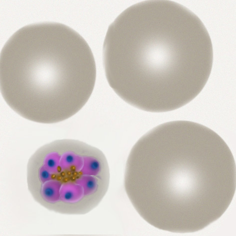
Brief summary
- Small rings (less delicate than P.falciparum) and becoming elongated or solid as parasites mature
- Red cells often small remaining a round shape and with no added dots unless heavily stained
- All forms tend to circulate, characteristically look for "daisy" schizonts and small round gametocytes
- Parasite number is often low
For more information
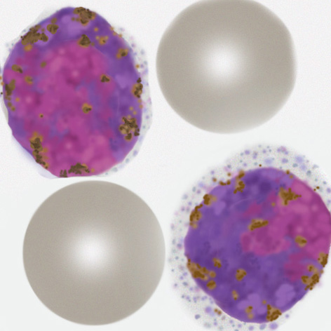
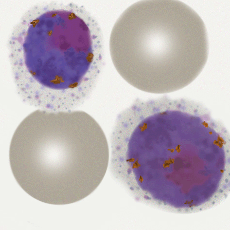
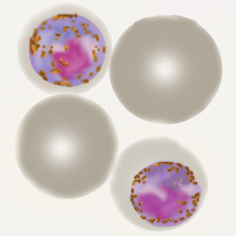
| Plasmodium knowlesi |
Early trophozoite
Late trophozoite
Schizont

Gametocyte
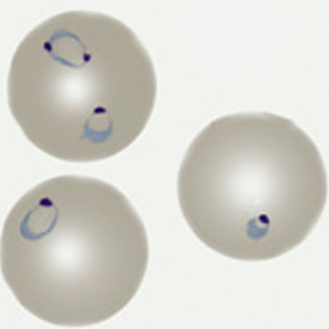

Brief Summary
- Very limited geographical distribution within S.E Asia
- Small fine ring forms resemble those of P.falciparum and may have high parasite count
- Later rings are more solid or elongated similar to P.malariae, although faint dots may be present
- Schizonts & gametocytes are often present and may resemble P.malariae but are less "neat"
- Characteristically red cell size is unaffected, although distortion may be seen
For more information
- click for full description of P.knowlesis morphology
- haga clic para obtener una descripción completa de la morfología de P.knowlesis
- click to visit the gallery of P.knowlesi forms