Schizont Development: Difference between revisions
From haematologyetc.co.uk
No edit summary |
No edit summary |
||
| Line 17: | Line 17: | ||
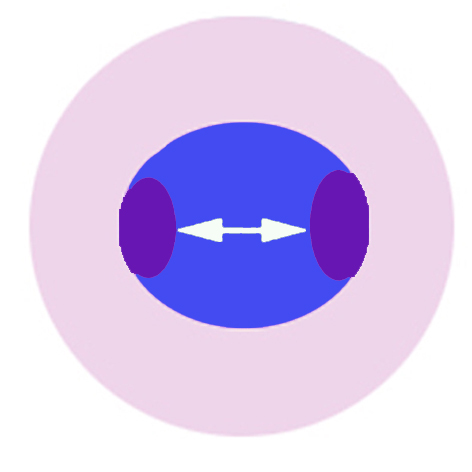
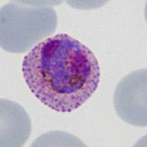
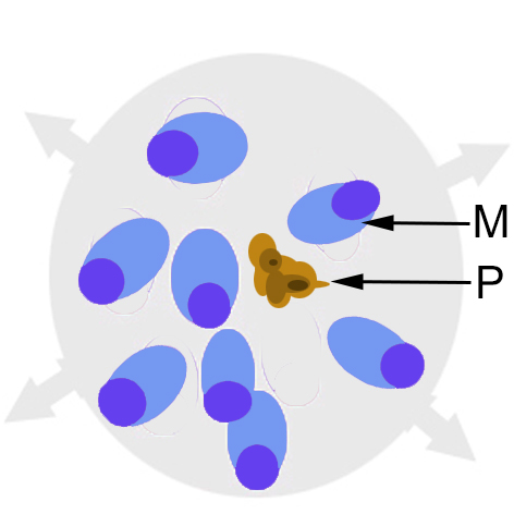
The first recognisable stage occurs when | The first recognisable stage occurs when the schizonts first divide their chromatin to form two distinct masses. This first stage is the least distinctive and can be difficult to distinguish from a late trophozoite or gametocyte with a double chromatin dot. But often the appearance is clear. | ||
<gallery mode="nolines" widths="200px" heights="220px" > | <gallery mode="nolines" widths="200px" heights="220px" > | ||
| Line 24: | Line 25: | ||
</gallery> | </gallery> | ||
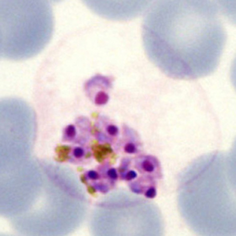
The cartoon image (A) shows the division of chromatin into two masses within a continuous blue parasite | The cartoon image (A) shows the division of chromatin into two masses within a continuous blue parasite cytoplasm (indiviual merozoites are not really distinguishable here). A clinical image of a parasite at this developmental stage (''P.ovale'' with well shown James'dots) is shown in panel (B). | ||
| Line 32: | Line 33: | ||
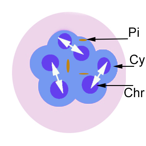
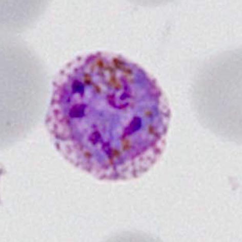
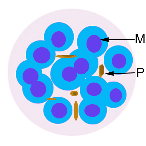
As schizont development proceeds further cycles of division cause the appearance of mutiple separate areas chromatin that will eventually form the merozoies, although at this stage they still lie within a single cytoplasmic mass. The number of divisions varies between species, so in mature schizonts this can contribute to species identification (see schizont gallery). | |||
<gallery mode="nolines" widths="200px" heights="220px" > | <gallery mode="nolines" widths="200px" heights="220px" > | ||
| Line 39: | Line 41: | ||
</gallery> | </gallery> | ||
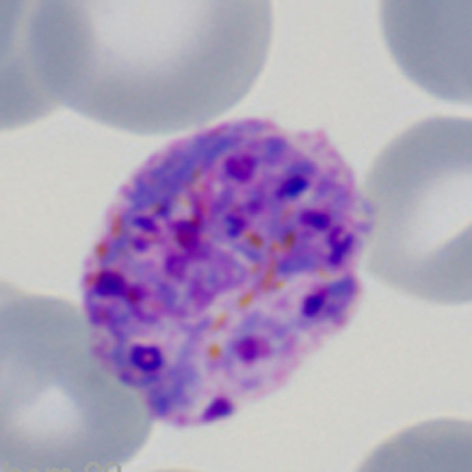
The cartoon image (A) shows the further division of chromatin into many discrete massed within the blue parasite cytoplasm (indiviual merozoites are still not distinguishable). A clinical image of a parasite at this developmental stage (again from ''P.ovale'' with well shown James'dots) is shown in panel (B). | |||
Revision as of 12:59, 27 March 2024
Navigation
Go Back
| How does schizont appearance change during their development?
THE INITIAL ASEXUAL DIVISION
The cartoon image (A) shows the division of chromatin into two masses within a continuous blue parasite cytoplasm (indiviual merozoites are not really distinguishable here). A clinical image of a parasite at this developmental stage (P.ovale with well shown James'dots) is shown in panel (B).
IMMATURE SCHIZONT APPEARANCES
The cartoon image (A) shows the further division of chromatin into many discrete massed within the blue parasite cytoplasm (indiviual merozoites are still not distinguishable). A clinical image of a parasite at this developmental stage (again from P.ovale with well shown James'dots) is shown in panel (B).
MATURE SCHIZONT APPEARANCES xxxxxx
xxxxxx.
xxxxxx
xxxxxx. |