| How does schizont appearance change during their development?
The schizonts we see on blood films are each at a particular stage of the successive cycles of asexual division that eventually result in the formation of multiple separate "merozoite" forms. Those merozoites are released as the red cell breaks down then go on to infect another red cell. Schizonts therefre look very different depending on which stage of development they represent. Below are images of schizonts at different developmental stages.
THE INITIAL ASEXUAL DIVISION
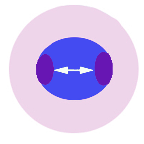
The first recognisable stage occurs when th schizonts first divide their chromatin to form two distinct masses. This first stage is the least distinctive and can be difficult t distinguish from a late trophozoite or gametocyte with a double chromatin dot. But often the appearance is clear.
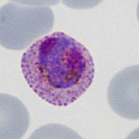
The cartoon image (A) shows the division of chromatin into two masses within a continuous blue parasite cytoplams (indiviual merozoites are not really distinguishable here). A clinical image of a parasite at this developmental stage (P.ovale with well shown James'dots) is shown in panel (B).
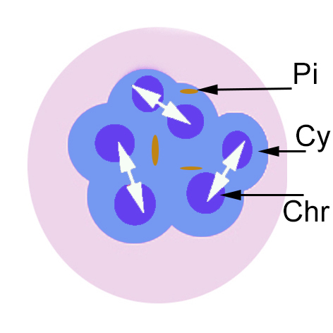
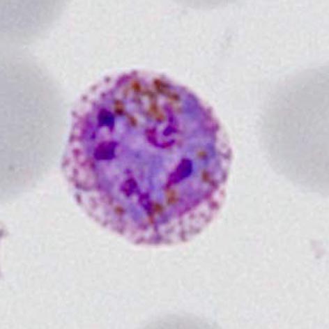
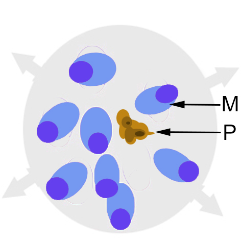
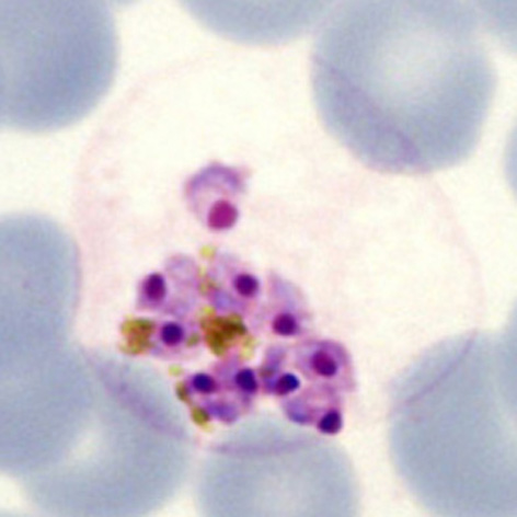
IMMATURE SCHIZONT APPEARANCES
xxxxxx
xxxxxx.
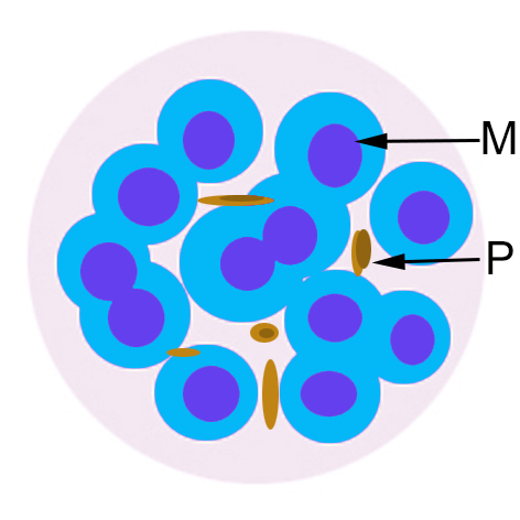
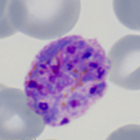
MATURE SCHIZONT APPEARANCES
xxxxxx
xxxxxx.
MEROZOITE RELEASE
xxxxxx
xxxxxx.
|