Schizont Development
From haematologyetc.co.uk
Navigation
Go Back
| How does schizont appearance change during their development?
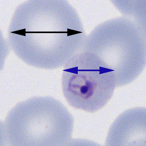
THE INITIAL ASEXUAL DEIVISION P.malariae The red cells in this species remain round and are often small in size
The early (A) and late trophozoites (B) shown in this image each lie within round erythrocytes with reduced size.
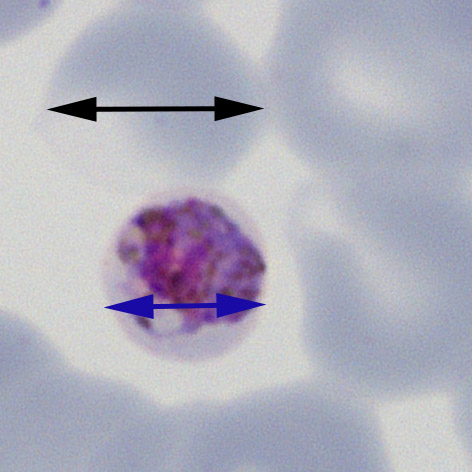
IMMATURE SCHIZONT APPEARANCES P.falciparum (and P.knowlesi) Red cell size and shape is generally unchanged although they may become crenated
The early (A) trophozoites lie within red cells that do not change size or shape, at later development (B) they may remain unchanged or acquire subtle crenation.
MATURE SCHIZONT APPEARANCES For both P.ovale and P.vivax the red cells become progressively enlarged and distorted as the parasites develop. It may not be possible to distingish the species based on red cell appearances, but there are differences which should be looked for.
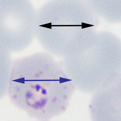
Expect increased red cell size but this may not be marked; the typical shape is an ovoid shape (hence the name) and there may be characteristic finbriation of cytoplams (that may be limited to one pole of the cell).
Early (A) and late (B) trophozoites o P.ovale. In each case there is a tendency for red cells to have an ovoid shape and there is distortion of the cytoplasm with sharp projectiosn (fimbriation). These orregular and spiky projections differ from the rounded crenation that may be seen in P.falciparum.
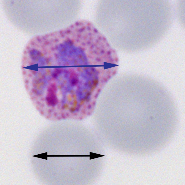
This species tend to have the largest red cell size that becomes evident at quite and early stage; the typical shape is quite irregular fimbriation is not (generally) seen.
Trophozoites of P.vivax cause increase in size and distortion of red cells as the parasites mature. Here, the the early trophozoite (A) is enlarged but still retains a relatively undistorted elongated shape (similar to P.ovale); however the late form (B) is has a very irregular shape (note that unlike P.ovale the red cell is not fimbriated). |