Schüffner's dots: Difference between revisions
From haematologyetc.co.uk
No edit summary |
No edit summary |
||
| (5 intermediate revisions by the same user not shown) | |||
| Line 12: | Line 12: | ||
<gallery mode="nolines" widths=250px heights=250px> | <gallery mode="nolines" widths=250px heights=250px> | ||
File: | File:Schuf2.jpg|link={{filepath:Schuf2.jpg}} | ||
</gallery> | </gallery> | ||
<span style="font-size: | |||
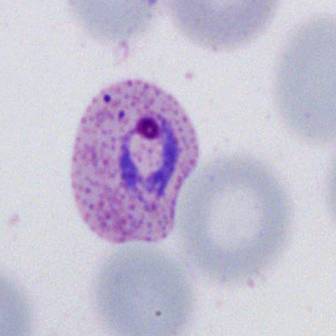
<span style="font-size:100%">Well formed Schüffner's dots in a trophozoite of ''P.vivax''; note that the dots are far too numerous to count and have a red/purple colour; remeber also that their appearance is highly dependent on the correct staining pH.</span> | |||
<br clear=all> | <br clear=all> | ||
| Line 21: | Line 22: | ||
<span style="color:navy>'''Species significance'''</span> | <span style="color:navy>'''Species significance'''</span> | ||
These dots are a feature of ''P.vivax'', but are morphologically indistinguishable from the James dots of ''P.ovale''. Distinction between these species must therefore be made based on other diagnostic crieria. | |||
---- | ---- | ||
| Line 28: | Line 30: | ||
<gallery mode="nolines" widths=200px heights=200px> | <gallery mode="nolines" widths=200px heights=200px> | ||
File: | File:Schuf1.jpg|A|link={{filepath:Schuf1.jpg}} | ||
File: | File:Schuf3.jpg|B|link={{filepath:Schuf3.jpg}} | ||
File: | File:Schuf4.jpg|C|link={{filepath:Schuf4.jpg}} | ||
</gallery> | </gallery> | ||
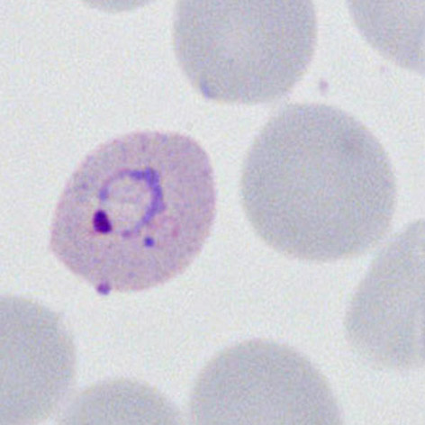
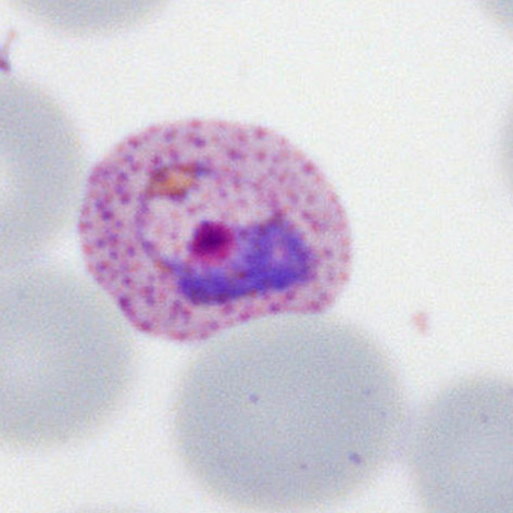
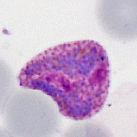
<span style="font-size: | <span style="font-size:100%">The appearance of Schüffner's dots during the develoment of ''P.vivax'' trophozites. These are shown from their earliest detectable form in an early trophozoite that has also begun to increase the size and distort the erythrocyte (A), these are more fully developed at later stages (B) and are highly visible in the amoeboid late trophozoite (C). </span> | ||
Latest revision as of 13:44, 3 April 2024
Navigation
Go Back
| What are Schüffner's dots?
Schüffner's dots are red-purple dots seen in P.vivax. They are morphologically indistinguishable from the James' dots of P.ovale but are very diffrent from the more dense and blue coloured Maurer's dots and clefts of P.falciparum. Like all parasite structures Schüffner's dots form progressively, and may not be seen in very early trophozoites.
Well formed Schüffner's dots in a trophozoite of P.vivax; note that the dots are far too numerous to count and have a red/purple colour; remeber also that their appearance is highly dependent on the correct staining pH.
Species significance These dots are a feature of P.vivax, but are morphologically indistinguishable from the James dots of P.ovale. Distinction between these species must therefore be made based on other diagnostic crieria.
Additional images
The appearance of Schüffner's dots during the develoment of P.vivax trophozites. These are shown from their earliest detectable form in an early trophozoite that has also begun to increase the size and distort the erythrocyte (A), these are more fully developed at later stages (B) and are highly visible in the amoeboid late trophozoite (C). |