Re-infection of the mosquito: Difference between revisions
From haematologyetc.co.uk
No edit summary |
No edit summary |
||
| (7 intermediate revisions by the same user not shown) | |||
| Line 9: | Line 9: | ||
{| class="wikitable" style="border-style: solid; border-width: 5px; border-color: #023020; color:black" | {| class="wikitable" style="border-style: solid; border-width: 5px; border-color: #023020; color:black" | ||
|colspan="1" style = "font-size:100%; color:black; background: #afbddb |''' | |colspan="1" style = "font-size:100%; color:black; background: #afbddb |'''Gametogenesis and sexual replication''' | ||
|} | |} | ||
<gallery mode="nolines" widths=300px heights=300px> | <gallery mode="nolines" widths=300px heights=300px> | ||
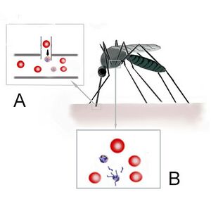
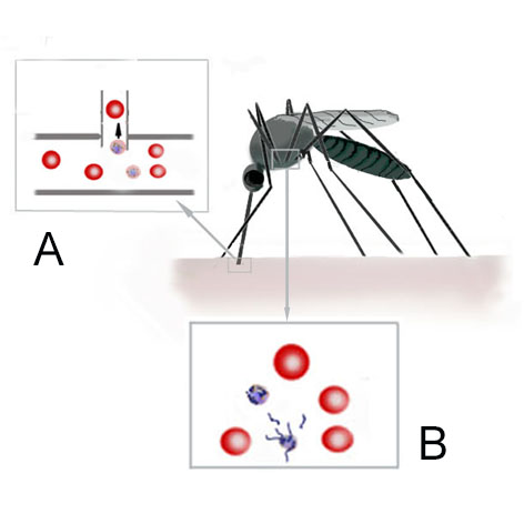
File:Mosquito uptake.jpg|<span style="font-size:90%">'' | File:Mosquito uptake.jpg|<span style="font-size:90%">''Development within the the mosquito''</span>|link={{filepath:Mosquito uptake.jpg}} | ||
</gallery> | |||
When a mosquito feeds from a malaria infected host, the blood meal will contain parasites of all forms. Importantly these forms include male and female gametocytes. The pH and temperature changes with the mosquito midgut cause the gametocytes to gain full sexual maturity (gametogenesis). The male gametocyte then undergoes 3 cycles of division to generate 8 male sexual forms (gametes). These then 8 gamaetes then "break out" of the male microgametocytes in a process known as exflagellation, and fuse with the female oocyte (derived from the macrogametocyte). Then following successful fusion move to the next stage of development with the mosquito gut wall. Finally the parasite enter the salivary glands of the mosquito as sporozoites where a new infection can be initiated. | |||
---- | |||
{| class="wikitable" style="border-style: solid; border-width: 5px; border-color: #023020; color:black" | |||
|colspan="1" style = "font-size:100%; color:black; background: #afbddb |'''Relevance to blood''' | |||
|} | |||
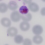
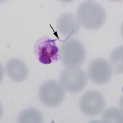
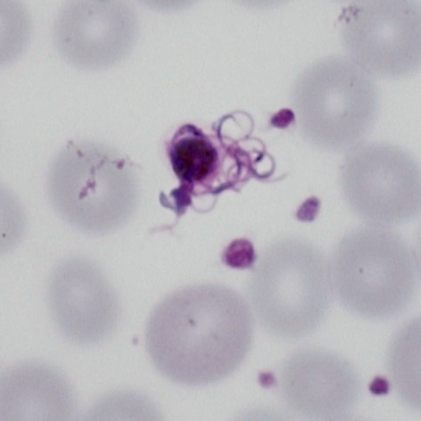
In stored samples of blood, the changes of storage may partly mimic the conditions within the mosquito midgut leading to changes that would only normally be seen in the mosquito. Some may be difficult to easily recognise, but are clearly atypical in appearance others have very clear appearances of exflagellation. It is important to be able to recognise these forms. | |||
<gallery mode="nolines" widths=180px heights=180px> | |||
File:ExFl1.jpg|<span style="font-size:80%">''A''</span>|link={{filepath:ExFl1.jpg}} | |||
File:ExFl2.jpg|<span style="font-size:80%">''B''</span>|link={{filepath:ExFl2.jpg}} | |||
File:ExFl3.jpg|<span style="font-size:80%">''C''</span>|link={{filepath:ExFl3.jpg}} | |||
File:ExFl4.jpg|<span style="font-size:80%">''D''</span>|link={{filepath:ExFl4.jpg}} | |||
</gallery> | </gallery> | ||
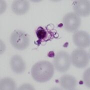
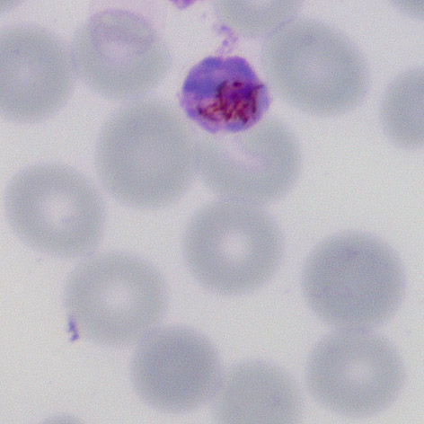
In the initial stage involves both the male microgametocytes and female macrogametocytes swelling and becoming more globular. The stage shown in (A) may represent an oocyte although it is not certain (these often clump together). The following stages are quite recognisable for the male gametocte as the male gametes burst from the erythrocytes in the process of exflagellation this may be seen as the early emergence (B), red cell swelling and disollution (C) and emergence of the gametes (D). | |||
---- | ---- | ||
Revision as of 14:24, 29 April 2024
Navigation
(click blue highlighted text to return to page)
Malaria main index
>Basic malaria biology
>>This page: Re-infection of the mosquito
| Gametogenesis and sexual replication |
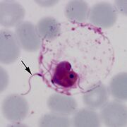
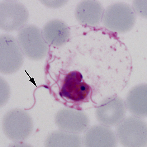
Development within the the mosquito
When a mosquito feeds from a malaria infected host, the blood meal will contain parasites of all forms. Importantly these forms include male and female gametocytes. The pH and temperature changes with the mosquito midgut cause the gametocytes to gain full sexual maturity (gametogenesis). The male gametocyte then undergoes 3 cycles of division to generate 8 male sexual forms (gametes). These then 8 gamaetes then "break out" of the male microgametocytes in a process known as exflagellation, and fuse with the female oocyte (derived from the macrogametocyte). Then following successful fusion move to the next stage of development with the mosquito gut wall. Finally the parasite enter the salivary glands of the mosquito as sporozoites where a new infection can be initiated.
| Relevance to blood |
In stored samples of blood, the changes of storage may partly mimic the conditions within the mosquito midgut leading to changes that would only normally be seen in the mosquito. Some may be difficult to easily recognise, but are clearly atypical in appearance others have very clear appearances of exflagellation. It is important to be able to recognise these forms.
A
B
C
D

In the initial stage involves both the male microgametocytes and female macrogametocytes swelling and becoming more globular. The stage shown in (A) may represent an oocyte although it is not certain (these often clump together). The following stages are quite recognisable for the male gametocte as the male gametes burst from the erythrocytes in the process of exflagellation this may be seen as the early emergence (B), red cell swelling and disollution (C) and emergence of the gametes (D).