P.vivx schizont gallery: Difference between revisions
From haematologyetc.co.uk
(Created page with "---- '''Navigation'''</br> Go Back ---- {| class="wikitable" style="border-style: solid; border-width: 4px; color:black" |colspan="1" style = "font-size:100%; color:black; background: FFFAFA"|<span style="color:black> {| class="wikitable" style="border-style: solid; border-width: 0px; border-color: #023020; color:black" |colspan="1" style = "font-size:100%; color:black; background: CBD5CO |'''''P.vivax'' gallery of late trophozoites'''''...") |
No edit summary |
||
| Line 18: | Line 18: | ||
<gallery mode="traditional" widths=240px heights=240px> | <gallery mode="traditional" widths=240px heights=240px> | ||
File:PVS1.jpg|<span style="font-size:80%">'''Developing late trophozoite''' The ring form is lost, the parasite is very irregular</span>|link={{filepath:PVS1.jpg}} | File:PVS1.jpg|<span style="font-size:80%">'''Developing late trophozoite''' The ring form is lost, the parasite is very irregular</span>|link={{filepath:PVS1.jpg}} | ||
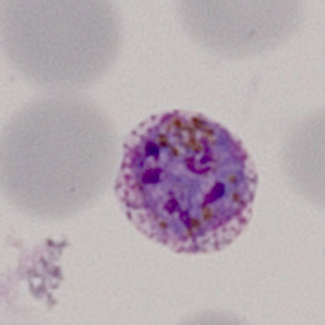
File: | File:PVS2.jpg|<span style="font-size:80%">'''Late trophozoite''' Note the solid parasite and the red cell begins to show distortion.</span>|link={{filepath:PVS3.jpg}} | ||
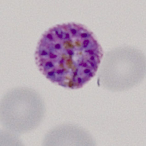
File: | File:PVS3.jpg|<span style="font-size:80%">'''Late trophozoite''' Similar to the previous example, the ring structure is entirely lost </span>|link={{filepath:PVS3.jpg}} | ||
</gallery>" | </gallery>" | ||
Latest revision as of 21:01, 7 April 2024
Navigation
Go Back
|