P.falciparum schizont gallery: Difference between revisions
From haematologyetc.co.uk
No edit summary |
No edit summary |
||
| (4 intermediate revisions by the same user not shown) | |||
| Line 12: | Line 12: | ||
<span style="font-size:95%">'''Summary'''</span> | <span style="font-size:95%">'''Summary'''</span> | ||
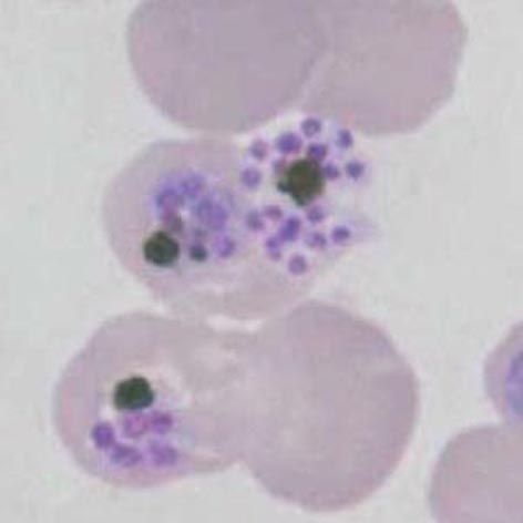
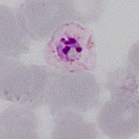
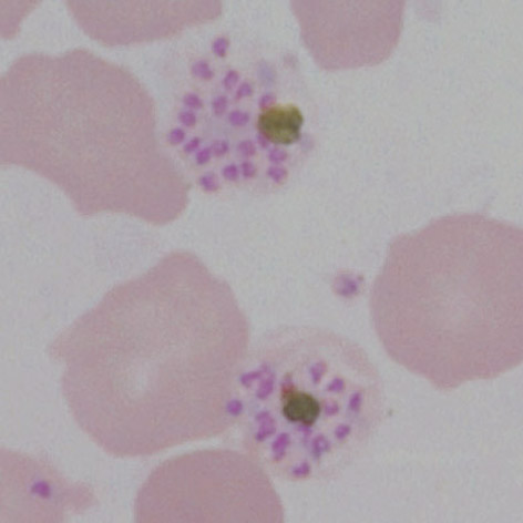
<span style="font-size:95%"> | <span style="font-size:95%">The schizonts of ''P.falciparum'' sequester in the small vessels and rarely circulate in blood; when found they usually signify a very severe infection. If this is not the case then it is appropriate to ask if there is a different species causing the infection. Generally these are loosely formed with variable numbers of merozoites and a single clump of pigment, they are not "neat" like the parasites of ''P.malariae''. | ||
---- | ---- | ||
<gallery mode="traditional" widths=240px heights=240px> | <gallery mode="traditional" widths=240px heights=240px> | ||
File:PFS1p.jpg|<span style="font-size:80%">''' | File:PFS1p.jpg|<span style="font-size:80%">'''Mature schizonts''' note the clumped brown pigment surrounded by loosely arranged merooites, some are early forms that are less well separated</span>|link={{filepath:PFS1p.jpg}} | ||
File:PFS2p.jpg|<span style="font-size:80%">''' | File:PFS2p.jpg|<span style="font-size:80%">'''A single merozoite''' the parasite is recognisable with just 4-5 merozoites and no pigment within a degenerate erythrocyte</span>|link={{filepath:PFS2p.jpg}} | ||
File:PFS3p.jpg|<span style="font-size:80%">''' | File:PFS3p.jpg|<span style="font-size:80%">'''Late schizonts''': 16-20 late merozoites prior to release are well formed and clearly separated with obvious pigment</span>|link={{filepath:PFS3p.jpg}} | ||
</gallery>" | </gallery>" | ||
Latest revision as of 10:48, 21 March 2024
Navigation
Go Back
|