P.falciparum late trophozoites gallery: Difference between revisions
From haematologyetc.co.uk
(Created page with "---- '''Navigation'''</br> Go Back ---- {| class="wikitable" style="border-style: solid; border-width: 4px; color:black" |colspan="1" style = "font-size:100%; color:black; background: FFFAFA"|<span style="color:black> {| class="wikitable" style="border-style: solid; border-width: 0px; border-color: #023020; color:black" |colspan="1" style = "font-size:100%; color:black; background: CBD5CO |'''''P.falciparum'' gallery of early tropho...") |
No edit summary |
||
| (5 intermediate revisions by the same user not shown) | |||
| Line 7: | Line 7: | ||
|colspan="1" style = "font-size:100%; color:black; background: FFFAFA"|<span style="color:black> | |colspan="1" style = "font-size:100%; color:black; background: FFFAFA"|<span style="color:black> | ||
{| class="wikitable" style="border-style: solid; border-width: 0px; border-color: #023020; color:black" | {| class="wikitable" style="border-style: solid; border-width: 0px; border-color: #023020; color:black" | ||
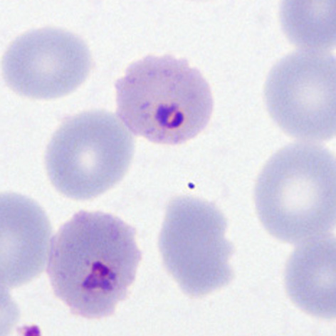
|colspan="1" style = "font-size:100%; color:black; background: CBD5CO |'''''P.falciparum'' gallery of | |colspan="1" style = "font-size:100%; color:black; background: CBD5CO |'''''P.falciparum'' gallery of late trophozoites''''' | ||
|} | |} | ||
<span style="font-size:95%">'''Summary'''</span> | <span style="font-size:95%">'''Summary'''</span> | ||
<span style="font-size:95%">At this stage we look for typical | <span style="font-size:95%">At this stage we look for rings that are slightly thicker though still small with typical ring form, the red cells tend to become crenated and pale, losing central pallor as the parasites mature. Typical accolé forms, double chromatin dot forms, and multiple parasites within infected red cells are still present. | ||
---- | ---- | ||
<gallery mode="traditional" widths=240px heights=240px> | <gallery mode="traditional" widths=240px heights=240px> | ||
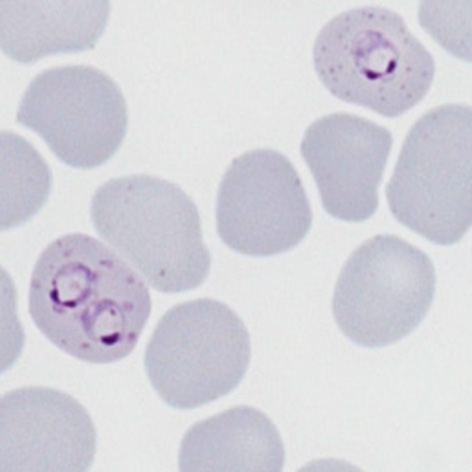
File:PFLT1p.jpg|<span style="font-size:80%">''' | File:PFLT1p.jpg|<span style="font-size:80%">'''Late rings''' Two cells both with typical dots: multiply infected and double dot forms</span>|link={{filepath:PFLT1p.jpg}} | ||
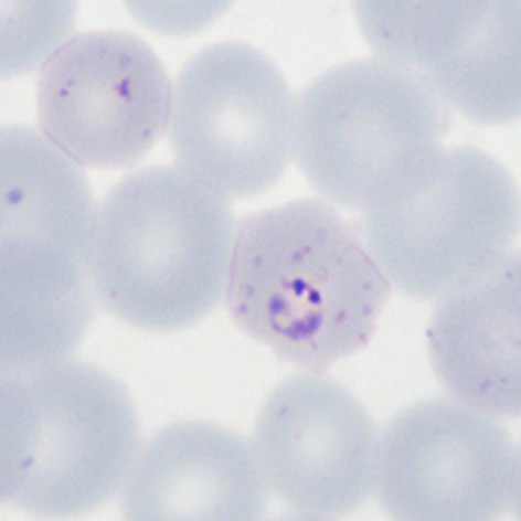
File:PFLT2p.jpg|<span style="font-size:80%">'''Double chromatin dot form''' | File:PFLT2p.jpg|<span style="font-size:80%">'''Double chromatin dot form''' also Maurers dost and clefts, slight crenation and lost pallor</span>|link={{filepath:PFLT2p.jpg}} | ||
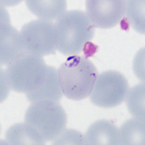
File:PFLT3p.jpg|<span style="font-size:80%">'''Accolé form''': | File:PFLT3p.jpg|<span style="font-size:80%">'''Accolé form''': closely associated with the red cell membrane, scanty mauers dots</span>|link={{filepath:PFLT3p.jpg}} | ||
File:PFLT4p.jpg|<span style="font-size:80%">''' | File:PFLT4p.jpg|<span style="font-size:80%">'''Accolé form''' A nice typical form with scanty well-formed Maurers dots</span>|link={{filepath:PFLT4p.jpg}} | ||
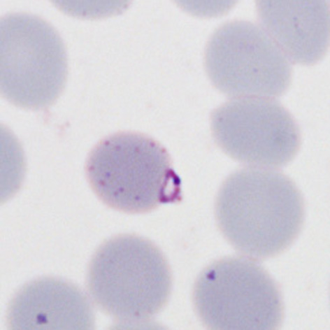
File:PFLT5p.jpg|<span style="font-size:80%">''' | File:PFLT5p.jpg|<span style="font-size:80%">'''Small thick forms''' the red cell crenation is well demonstrated with scanty dots</span>|link={{filepath:PFLT5p.jpg}} | ||
</gallery>" | </gallery>" | ||
Latest revision as of 00:30, 21 March 2024
Navigation
Go Back
|