P.falciparum gametocyte gallery: Difference between revisions
From haematologyetc.co.uk
No edit summary |
No edit summary |
||
| Line 16: | Line 16: | ||
---- | ---- | ||
<gallery mode="traditional" widths=240px heights=240px> | <gallery mode="traditional" widths=240px heights=240px> | ||
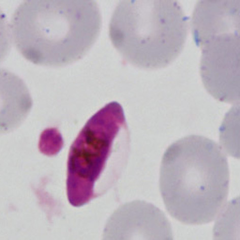
File:PFG2.jpg|<span style="font-size:80%">'''Macrogametocyte''' | File:PFG2.jpg|<span style="font-size:80%">'''Macrogametocyte''' A long rod with empty red cell membrane to the right side. The form is not very curved but the disortion of the rod is clearly beginning</span>|link={{filepath:PFG2.jpg}} | ||
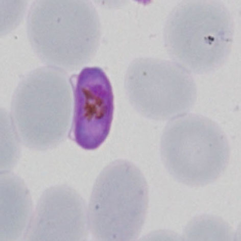
File:PFG3.jpg|<span style="font-size:80%">'''Microgametocyte''': the small rod does not fully fill the erythrocyte, the residual red cell membrane is lose to the side of the membrane</span>|link={{filepath:PFG3.jpg}} | File:PFG3.jpg|<span style="font-size:80%">'''Microgametocyte''': the small rod does not fully fill the erythrocyte, the residual red cell membrane is lose to the side of the membrane</span>|link={{filepath:PFG3.jpg}} | ||
File:PFG4.jpg|<span style="font-size:80%">'''Macrogametocyte''' A nice typical form | File:PFG4.jpg|<span style="font-size:80%">'''Macrogametocyte''' A nice typical "banana" form, the shape is very curved and pigment is seen scattered over he chromatin</span>|link={{filepath:PFG4.jpg}} | ||
</gallery>" | </gallery>" | ||
Latest revision as of 11:01, 21 March 2024
Navigation
Go Back
|