P.falciparum gametocyte gallery: Difference between revisions
From haematologyetc.co.uk
No edit summary |
No edit summary |
||
| (2 intermediate revisions by the same user not shown) | |||
| Line 12: | Line 12: | ||
<span style="font-size:95%">'''Summary'''</span> | <span style="font-size:95%">'''Summary'''</span> | ||
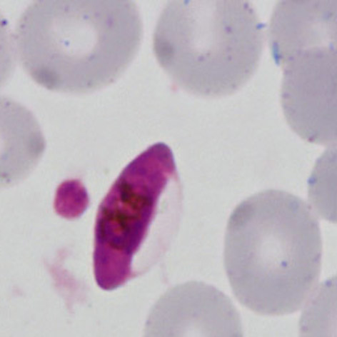
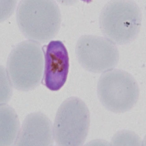
<span style="font-size:95%">Gametocytes in this species are highly distinctive. They develop within the red cell as long rods, however the red cell membrane continues to restrict them - the haemoglobin is fully metabolised so is not visible, but the residual membrane of the red cell "ghost" can usually be seen to the side of the red cell. The gametocytes differ with large (macrogametocyte) forms that become curved because of the red cell membrane restricting thir shape (banana form); the smaller (microgametocytes) retain the rod shape. | <span style="font-size:95%">Gametocytes in this species are highly distinctive. They develop within the red cell as long rods, however the red cell membrane continues to restrict them - the haemoglobin is fully metabolised so is not visible, but the residual membrane of the red cell "ghost" can usually be seen to the side of the red cell. The gametocytes differ with large (macrogametocyte) forms that become curved because of the red cell membrane restricting thir shape (banana form); the smaller (microgametocytes) retain the rod shape. In each case the gold/brown pigment overlies the chromatin area | ||
---- | ---- | ||
<gallery mode="traditional" widths=240px heights=240px> | <gallery mode="traditional" widths=240px heights=240px> | ||
File:PFG2.jpg|<span style="font-size:80%">''' | File:PFG2.jpg|<span style="font-size:80%">'''Macrogametocyte''' A long rod with empty red cell membrane to the right side. The form is not very curved but the disortion of the rod is clearly beginning</span>|link={{filepath:PFG2.jpg}} | ||
File:PFG3.jpg|<span style="font-size:80%">''' | File:PFG3.jpg|<span style="font-size:80%">'''Microgametocyte''': the small rod does not fully fill the erythrocyte, the residual red cell membrane is lose to the side of the membrane</span>|link={{filepath:PFG3.jpg}} | ||
File:PFG4.jpg|<span style="font-size:80%">''' | File:PFG4.jpg|<span style="font-size:80%">'''Macrogametocyte''' A nice typical "banana" form, the shape is very curved and pigment is seen scattered over he chromatin</span>|link={{filepath:PFG4.jpg}} | ||
</gallery>" | </gallery>" | ||
Latest revision as of 11:01, 21 March 2024
Navigation
Go Back
|