P.falciparum early trophozoites gallery: Difference between revisions
From haematologyetc.co.uk
No edit summary |
No edit summary |
||
| Line 20: | Line 20: | ||
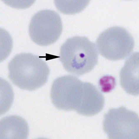
File:PFET3p.jpg|<span style="font-size:80%">'''Accolé form''': The arrowed form is closely associated with the red cell membrane</span>|link={{filepath:PFET3p.jpg}} | File:PFET3p.jpg|<span style="font-size:80%">'''Accolé form''': The arrowed form is closely associated with the red cell membrane</span>|link={{filepath:PFET3p.jpg}} | ||
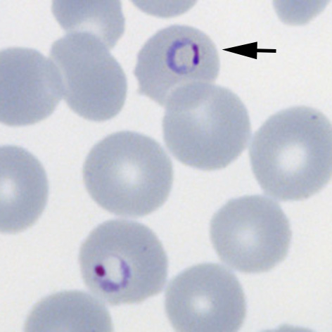
File:PFET4p.jpg|<span style="font-size:80%">'''Multiple parasites''' Two parasites within a single red cells (arrowed)</span>|link={{filepath:PFET4p.jpg}} | File:PFET4p.jpg|<span style="font-size:80%">'''Multiple parasites''' Two parasites within a single red cells (arrowed)</span>|link={{filepath:PFET4p.jpg}} | ||
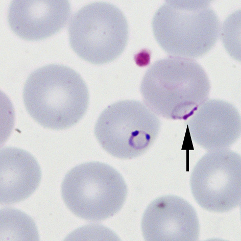
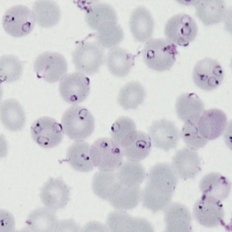
File:PFET5p.jpg|<span style="font-size:80%">'''High parasitaemia''' Most of the typical ''P.falciparum'' forms are present</span>|link={{filepath:PFET5p.jpg}} | File:PFET5p.jpg|<span style="font-size:80%">'''High parasitaemia''' Most of the typical early trophozoite ''P.falciparum'' forms are present</span>|link={{filepath:PFET5p.jpg}} | ||
</gallery>" | </gallery>" | ||
Revision as of 12:17, 20 March 2024
Navigation
Go Back
|