Biology of the schizont: Difference between revisions
From haematologyetc.co.uk
No edit summary |
No edit summary |
||
| (29 intermediate revisions by the same user not shown) | |||
| Line 1: | Line 1: | ||
{{DISPLAYTITLE:<span style="position: absolute; clip: rect(1px 1px 1px 1px); clip: rect(1px, 1px, 1px, 1px);">{{FULLPAGENAME}}</span>}} | |||
---- | ---- | ||
'''Navigation'''</br> | '''Navigation'''</br> | ||
<span style="font-size: | <span style="font-size:90%">>[[Malaria_Index|Main Malaria Index]]''</span></br> | ||
<span style="font-size:90%">[[Malaria Index | <span style="font-size:90%">>>[[Malaria_Biology|Malaria Biology Index]]''</span></br> | ||
<span style="font-size:90%">> | <span style="font-size:90%">>>>Current page: '''Schizont Biology'''</span> | ||
<span style="font-size: | |||
---- | |||
<span style="font-size:160%; color:navy">Biology of the Schizont</br></span> | |||
---- | ---- | ||
{| class="wikitable" style="border-style: solid; border-width: | {| class="wikitable" style="border-style: solid; border-width: 4px; border-color:light gray" | ||
|colspan="1" style = "font-size:100%; color:black; background: | |colspan="1" style = "font-size:100%; color:black; background: white"|<span style="color:navy></span> | ||
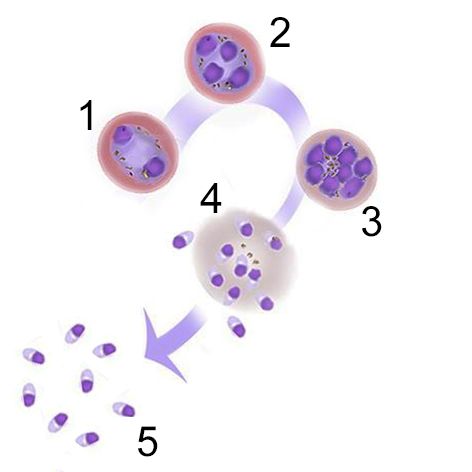
The stage begins with the first cycle of '''asexual replication''' forming a recognisable “schizont” then concludes when the individual “merozoites” are released to infect new erythrocytes. | The stage begins with the first cycle of '''asexual replication''' forming a recognisable “schizont” then concludes when the individual “merozoites” are released to infect new erythrocytes. | ||
<gallery mode="nolines" widths=300px heights=300px> | <gallery mode="nolines" widths=300px heights=300px> | ||
| Line 38: | Line 29: | ||
---- | ---- | ||
{| class="wikitable" style="border-style: | {| class="wikitable" style="border-style: none; border-width: 2px; border-color: gainsboro; color:black" | ||
|colspan="1" style = "font-size:100%; color:black; background: | |colspan="1" style = "font-size:100%; color:black; background: gainsboro|'''Morphological features and relevance''' | ||
|} | |} | ||
| Line 45: | Line 36: | ||
(1) '''The number of replication cycles differs between species:''' the typical number of merozoites formed differs between species with as few as 8 (in P.malariae) up to a possible 32 (in P.vivax)</br> | (1) '''The number of replication cycles differs between species:''' the typical number of merozoites formed differs between species with as few as 8 (in P.malariae) up to a possible 32 (in P.vivax)</br> | ||
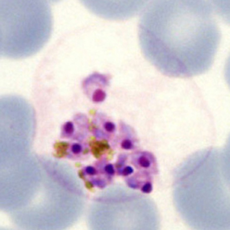
(2) '''This stage may not always occur in blood:''' schizonts of ''P.falciparum'' adhere within the small vessels so is not seen in blood unless infection is very severe | (2) '''This stage may not always occur in blood:''' schizonts of ''P.falciparum'' adhere within the small vessels so is not seen in blood unless infection is very severe | ||
</br> | </br></br> | ||
<gallery mode="nolines" widths="200px" heights="220px" > | |||
File:Schizontreal4.jpg|Mature schizont releasing merozoites|link={{filepath:Schizontreal4.jpg}} | |||
</gallery> | |||
<span style="font-size:10%"></span> The progressive maturation of this parasite stage means that they have a wide range of morphological forms. However, these can be readily recognised on blood films by reference to their biology | |||
[ [[Images of schizont morphology|See clinical images illustrating schizont development]] ] | |||
---- | ---- | ||
{| class="wikitable" style="border-style: | {| class="wikitable" style="border-style: none; border-width: 2px; border-color: gainsboro; color:black" | ||
|colspan="1" style = "font-size:100%; color:black; background: | |colspan="1" style = "font-size:100%; color:black; background: gainsboro |'''Relevance of schizonts to clinical biology''' | ||
|} | |} | ||
Latest revision as of 18:02, 6 November 2024
Navigation
>Main Malaria Index
>>Malaria Biology Index
>>>Current page: Schizont Biology
Biology of the Schizont
|
The stage begins with the first cycle of asexual replication forming a recognisable “schizont” then concludes when the individual “merozoites” are released to infect new erythrocytes.
The progressive maturation of this parasite stage means that they have a wide range of morphological forms. However, these can be readily recognised on blood films by reference to their biology [ See clinical images illustrating schizont development ]
|