Red cell crenation description: Difference between revisions
From haematologyetc.co.uk
(Created page with "---- '''Navigation'''</br> Go Back ---- {| class="wikitable" style="border-style: solid; border-width: 5px; color:black" |colspan="1" style = "font-size:100%; color:black; background: WhiteSmoke"|<span style="color:navy>'''When does red cell crenation occur?'''</span> This refers to crenation of erythrocytes that contain parasites when the normal erythrocytes are unaffected, i.e. it is a feature of the presence of parasites. The finding is so...") |
No edit summary |
||
| (One intermediate revision by the same user not shown) | |||
| Line 7: | Line 7: | ||
|colspan="1" style = "font-size:100%; color:black; background: WhiteSmoke"|<span style="color:navy>'''When does red cell crenation occur?'''</span> | |colspan="1" style = "font-size:100%; color:black; background: WhiteSmoke"|<span style="color:navy>'''When does red cell crenation occur?'''</span> | ||
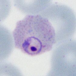
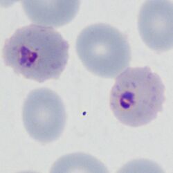
This refers to crenation of erythrocytes that contain parasites when the normal erythrocytes are unaffected, i.e. it is a feature of the presence of parasites. The finding is sometimes (though not always | This refers to crenation of erythrocytes that contain parasites when the normal erythrocytes are unaffected, i.e. it is a feature of the presence of parasites. The finding is sometimes (though not always) seen in ''P.falciparum'' infection, particularly in later forms, and presumably rreflects altered red cell hydration. | ||
<gallery mode="nolines" widths=250px heights=250px> | <gallery mode="nolines" widths=250px heights=250px> | ||
File: | File:MFcrenation1.jpg|link={{filepath:MFcrenation1.jpg}} | ||
File:MFcrenation2.jpg|link={{filepath:MFcrenation2.jpg}} | |||
</gallery> | </gallery> | ||
<span style="font-size:80%">Note that the parasite is very closely in contact with the red cell membrane (''P.falciparum'' late trophozoite form with maurer's dots and clefts)</span> | <span style="font-size:80%">Note that the parasite is very closely in contact with the red cell membrane (''P.falciparum'' late trophozoite form with maurer's dots and clefts)</span> | ||
| Line 20: | Line 21: | ||
<span style="color:navy>'''Species significance'''</span> | <span style="color:navy>'''Species significance'''</span> | ||
Although most frequently a feature of ''P.falciparum'' infection, the feature is often not seen, and should not be considered fully species-specific. | |||
---- | ---- | ||
Latest revision as of 18:35, 6 June 2024
Navigation
Go Back
| When does red cell crenation occur?
This refers to crenation of erythrocytes that contain parasites when the normal erythrocytes are unaffected, i.e. it is a feature of the presence of parasites. The finding is sometimes (though not always) seen in P.falciparum infection, particularly in later forms, and presumably rreflects altered red cell hydration.
Note that the parasite is very closely in contact with the red cell membrane (P.falciparum late trophozoite form with maurer's dots and clefts)
Species significance Although most frequently a feature of P.falciparum infection, the feature is often not seen, and should not be considered fully species-specific. |

