Stained correctly: Difference between revisions
From haematologyetc.co.uk
(Created page with "description staining: more alkaline than normal films is recommended for malaria parasite evaluation - irrespective of colour preference this makes parasites more visible compared with the grey of the surrounding red cells and makes some structures such as cytoplasmic dots more visible.") |
No edit summary |
||
| Line 1: | Line 1: | ||
---- | |||
'''Navigation'''</br> | |||
[[Plasmodium falciparum: Morphology|Go Back]] | |||
---- | |||
staining | {| class="wikitable" style="border-style: solid; border-width: 4px; color:black" | ||
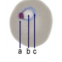
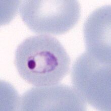
|colspan="1" style = "font-size:100%; color:black; background: FFFAFA"|<span style="color:navy>'''What is a ring form?'''</span> | |||
<gallery mode="nolines" widths="220px" heights="220px" > | |||
File:Ring_c.jpg|link={{filepath:|Ring_c.jpg}} | |||
File:MMETr.jpg|link={{filepath:|MMETr.jpg}} | |||
</gallery> | |||
The earliest stage following red cell invasion has a typical ting form: | |||
---- | |||
<span style="color:navy>'''Description'''</span> | |||
The best detection and species identification in malaria is made when staining is performed at a slightly more alkaline pH - this makes parasites more visible compared with the grey of the surrounding red cells and makes some structures such as cytoplasmic dots more visible. These features are detectable at standard staining pH and both detection and species identification can be performed, it is just less easy. | |||
Revision as of 11:07, 24 March 2024
Navigation
Go Back
| What is a ring form?
The earliest stage following red cell invasion has a typical ting form: Description
|