Species identification: summary page: Difference between revisions
From haematologyetc.co.uk
No edit summary |
No edit summary |
||
| (67 intermediate revisions by 2 users not shown) | |||
| Line 1: | Line 1: | ||
---- | ---- | ||
'''Navigation'''</br> | '''Navigation'''</br> | ||
<span style="font-size:80%">(click blue highlighted text to return to page)</span></br></br> | |||
<span style="font-size:90%">[[Malaria Index|Malaria main index]]''</span></br> | <span style="font-size:90%">[[Malaria Index|Malaria main index]]''</span></br> | ||
<span style="font-size:90%">>This page: <u>Species Indentification: summary</u></span> | <span style="font-size:90%">>This page: <u>Species Indentification: summary</u></span> | ||
---- | ---- | ||
{| class="wikitable" style="border-style: none; border-width: 5px; border-color: #023020; color:black" | |||
|colspan="1" style = "font-size:100%; color:black; background: #afbddb |'''''Plasmodium falciparum''''' | |||
|} | |||
<gallery mode="nolines" widths= | <gallery mode="nolines" widths=140px heights=150px> | ||
File:PFETc.jpg|<span style="font-size:80%">''Early trophozoite''</span>|link={{filepath:PFETc.jpg}} | File:PFETc.jpg|<span style="font-size:80%">''Early trophozoite''</span>|link={{filepath:PFETc.jpg}} | ||
File:PFLTc.jpg|<span style="font-size:80%">Late trophozoite</span>|link={{filepath:PFLTc.jpg}} | File:PFLTc.jpg|<span style="font-size:80%">Late trophozoite</span>|link={{filepath:PFLTc.jpg}} | ||
File: | File:PFSc2.jpg|<span style="font-size:80%">Schizont (rare)</span>|link={{filepath:PFSc2.jpg}} | ||
File:PFGc.jpg|<span style="font-size:80%">Gametocyte</span>|link={{filepath:PFGc.jpg}} | File:PFGc.jpg|<span style="font-size:80%">Gametocyte</span>|link={{filepath:PFGc.jpg}} | ||
</gallery> | </gallery> | ||
<span style="font-size: | <span style="font-size:90%">'''Summary:''' Trophozoites are small, fine "rings", typically with accolé forms, multiple parasites per cell, and double dot forms. At later developmental stages trophozoites aquire characteristic Maurer's dots and clefts. Schizonts are irregular and "tatty" but are rarely seen in blood unless severe infection. The gametocytes have a characteristic elongated (often curved) 'banana' form.</span> | ||
<div style="width: 230px"> | |||
{| class="wikitable" style="border-left:solid 4px gray;border-right:solid 4px gray;border-top:solid 4px gray;border-bottom:solid 4px gray; font-size:90%; color:navy; align:center" | |||
<div style="width: | |||
{| class="wikitable" style="border-left:solid 4px | |||
| colspan="1"''|[[Plasmodium falciparum: Morphology|'''CLICK''' for detailed description]]'' | | colspan="1"''|[[Plasmodium falciparum: Morphology|'''CLICK''' for detailed description]]'' | ||
|} | |} | ||
| Line 31: | Line 28: | ||
---- | ---- | ||
<div style="width: 95%"> | <div style="width: 95%"> | ||
{| class="wikitable" style="border-style: solid; border-width: 5px; color:black" | {| class="wikitable" style="border-style: solid; border-width: 5px; border-color: #023020; color:black" | ||
|colspan="1" style = "font-size: | |colspan="1" style = "font-size:100%; color:black; background: #afbddb |'''''Plasmodium vivax''''' | ||
|} | |} | ||
<gallery mode="nolines" widths=140px heights=140px> | |||
File:PVETc.jpg|<span style="font-size:80%">''Early trophozoite''</span>|link={{filepath:PVETc.jpg}} | |||
File:PVLTc.jpg|<span style="font-size:80%">Late trophozoite</span>|link={{filepath:PVLTc.jpg}} | |||
File:PVSc.jpg|<span style="font-size:80%">Schizont (rare)</span>|link={{filepath:PVSc.jpg}} | |||
File:PVGc.jpg|<span style="font-size:80%">Gametocyte</span>|link={{filepath:PVGc.jpg}} | |||
</gallery> | |||
''' | '''Summary''' | ||
*Large and robust rings that become amoeboid during later development | *Large and robust rings that become "amoeboid" during later development | ||
*Red cells become increasingly enlarged and distorted as parasites mature | *Red cells become increasingly enlarged and distorted as parasites mature | ||
*Schüffner's dots are visible in appropriately stained thin blood films | *Schüffner's dots are visible in appropriately stained thin blood films | ||
| Line 57: | Line 52: | ||
'' | <div style="width: 230px"> | ||
{| class="wikitable" style="border-left:solid 4px gray;border-right:solid 4px gray;border-top:solid 4px gray;border-bottom:solid 4px gray; font-size:90%; color:navy; align:center" | |||
| colspan="1"''|[[Plasmodium vivax: Morphology|'''CLICK''' for detailed description]]'' | |||
|} | |||
</div> | |||
---- | ---- | ||
<div style="width: 95%"> | <div style="width: 95%"> | ||
{| class="wikitable" style="border-style: solid; border-width: 5px; color:black" | {| class="wikitable" style="border-style: solid; border-width: 5px; border-color: #023020; color:black" | ||
|colspan="1" style = "font-size: | |colspan="1" style = "font-size:100%; color:black; background: #afbddb |'''''Plasmodium ovale''''' | ||
|} | |} | ||
[[File:POETc.jpg|150px|link={{filepath:POETc.jpg}}]] | [[File:POETc.jpg|150px|link={{filepath:POETc.jpg}}]] | ||
[[File:POLTc.jpg|150px|link={{filepath:POLTc.jpg}}]] | [[File:POLTc.jpg|150px|link={{filepath:POLTc.jpg}}]] | ||
| Line 76: | Line 77: | ||
'''Brief summary''' | '''Brief summary''' | ||
* | *Ring forms are large and robust and often retained in the late trophozoite stage | ||
*Red cells | *Red cells become moderately enlarged and may have oval shape with characteristic fimbriation | ||
*Schüffner's (James) dots seen in appropriately stained samples | *Schüffner's (James) dots form dusing development and will be seen in appropriately stained samples | ||
*All forms tend to circulate | *All developmental forms tend to circulate and may be difficult to distinguish from ''P.vivax'' | ||
'' | <div style="width: 230px"> | ||
{| class="wikitable" style="border-left:solid 4px gray;border-right:solid 4px gray;border-top:solid 4px gray;border-bottom:solid 4px gray; font-size:90%; color:navy; align:center" | |||
| colspan="1"''|[[Plasmodium ovale: Morphology|'''CLICK''' for detailed description]]'' | |||
|} | |||
</div> | |||
---- | ---- | ||
<div style="width: 95%"> | <div style="width: 95%"> | ||
{| class="wikitable" style="border-style: solid; border-width: 5px; color:black" | {| class="wikitable" style="border-style: solid; border-width: 5px; border-color: #023020; color:black" | ||
|colspan="1" style = "font-size: | |colspan="1" style = "font-size:100%; color:black; background: #afbddb |'''''Plasmodium malariae''''' | ||
|} | |} | ||
[[File:PMETc.jpg|150px|link={{filepath:PMETc.jpg}}]] | [[File:PMETc.jpg|150px|link={{filepath:PMETc.jpg}}]] | ||
[[File:PMLTc.jpg|150px|link={{filepath:PMLTc.jpg}}]] | [[File:PMLTc.jpg|150px|link={{filepath:PMLTc.jpg}}]] | ||
| Line 113: | Line 118: | ||
---- | ---- | ||
{| class="wikitable" style="border-style: solid; border-width: 5px; color:black" | {| class="wikitable" style="border-style: solid; border-width: 5px; border-color: #023020; color:black" | ||
|colspan="1" style = "font-size: | |colspan="1" style = "font-size:100%; color:black; background: #fff5ee |'''''Plasmodium knowlesi''''' | ||
|} | |} | ||
<gallery mode="nolines" widths=150px heights=150px> | <gallery mode="nolines" widths=150px heights=150px> | ||
File:PKETc.jpg|<span style="font-size:80%">''Early trophozoite''</span>|link={{filepath:PKETc.jpg}} | File:PKETc.jpg|<span style="font-size:80%">''Early trophozoite''</span>|link={{filepath:PKETc.jpg}} | ||
| Line 137: | Line 143: | ||
''For more information'' | ''For more information'' | ||
*[[''Plasmodium knowlesi'': Morphology|click for full description of ''P.knowlesis'' morphology]] | *[[''Plasmodium knowlesi'': Morphology|click for full description of ''P.knowlesis'' morphology]] | ||
*[[''Plasmodium knowlesi'': Morfología|haga clic para obtener una descripción completa de la morfología de ''P.knowlesis'']] | |||
*[[''Plasmodium knowlesi'': Gallery|click to visit the gallery of ''P.knowlesi'' forms]] | *[[''Plasmodium knowlesi'': Gallery|click to visit the gallery of ''P.knowlesi'' forms]] | ||
---- | ---- | ||
Latest revision as of 13:40, 20 May 2024
Navigation
(click blue highlighted text to return to page)
Malaria main index
>This page: Species Indentification: summary
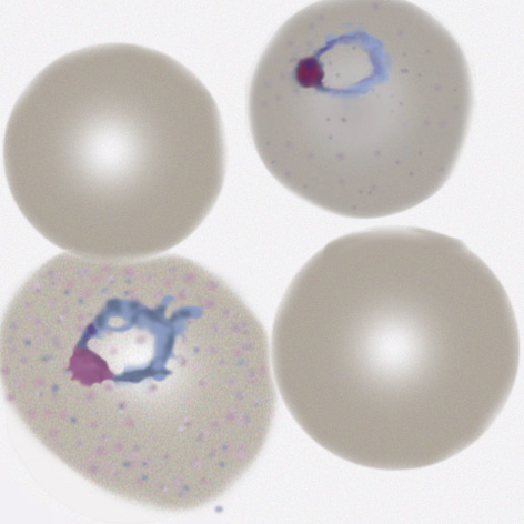
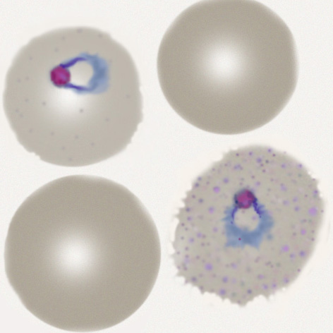
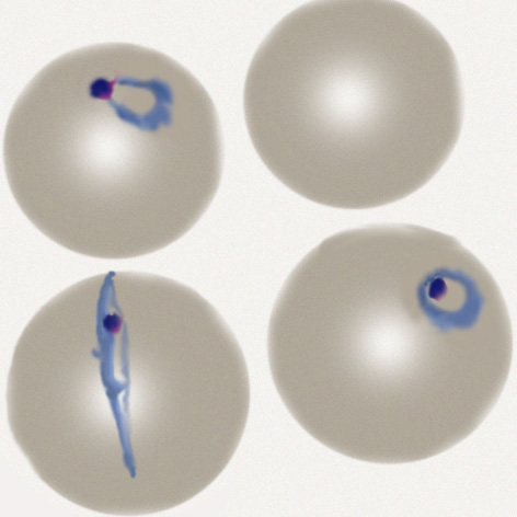
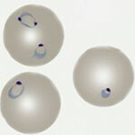
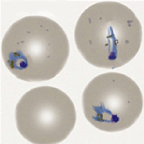
| Plasmodium falciparum |
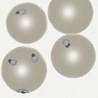
Early trophozoite
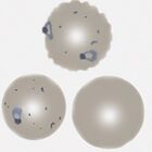
Late trophozoite

Schizont (rare)
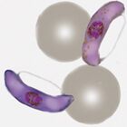
Gametocyte

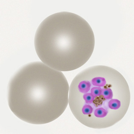

Summary: Trophozoites are small, fine "rings", typically with accolé forms, multiple parasites per cell, and double dot forms. At later developmental stages trophozoites aquire characteristic Maurer's dots and clefts. Schizonts are irregular and "tatty" but are rarely seen in blood unless severe infection. The gametocytes have a characteristic elongated (often curved) 'banana' form.
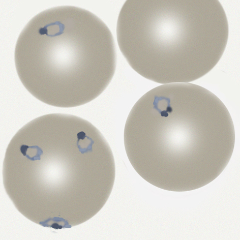
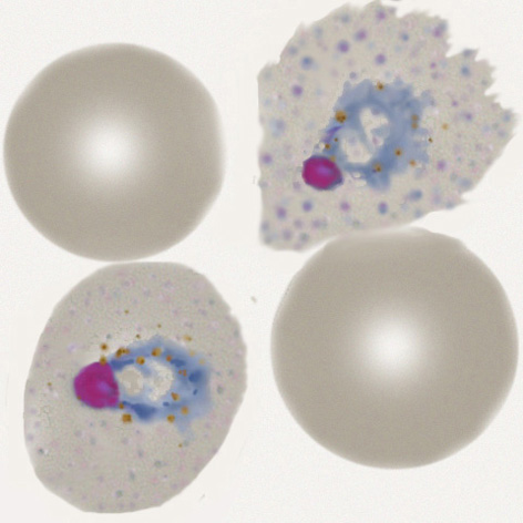
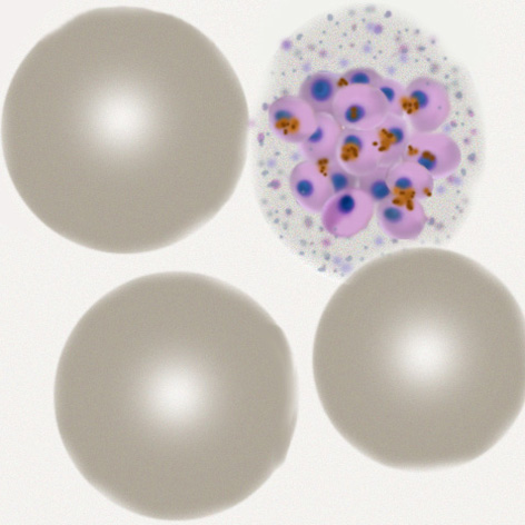
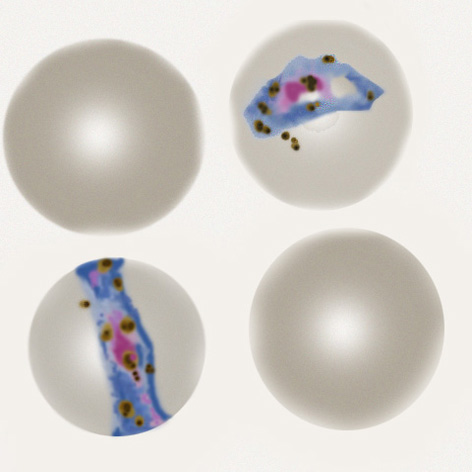
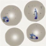
| Plasmodium vivax |
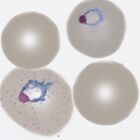
Early trophozoite
Late trophozoite

Schizont (rare)
Gametocyte
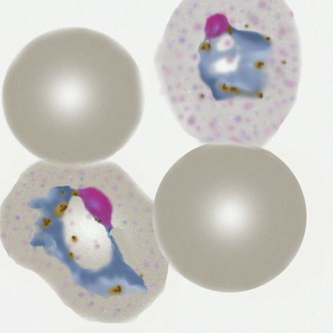
Summary
- Large and robust rings that become "amoeboid" during later development
- Red cells become increasingly enlarged and distorted as parasites mature
- Schüffner's dots are visible in appropriately stained thin blood films
- All forms tend to circulate with large schizont and gametocyte forms present
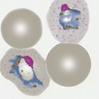
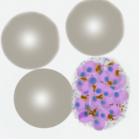
| Plasmodium ovale |
Brief summary
- Ring forms are large and robust and often retained in the late trophozoite stage
- Red cells become moderately enlarged and may have oval shape with characteristic fimbriation
- Schüffner's (James) dots form dusing development and will be seen in appropriately stained samples
- All developmental forms tend to circulate and may be difficult to distinguish from P.vivax
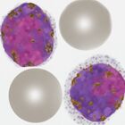
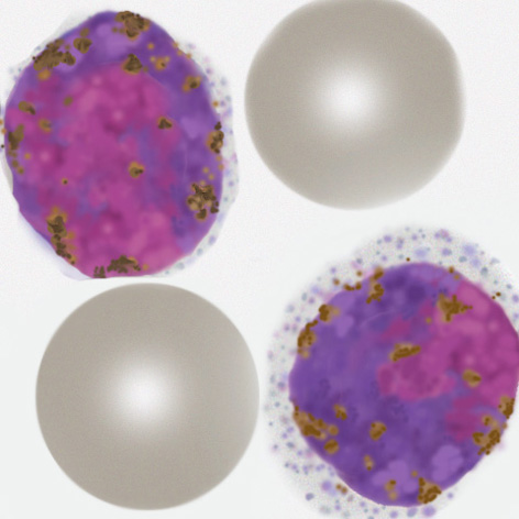
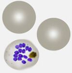
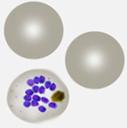
| Plasmodium malariae |
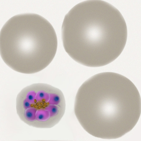
Brief summary
- Small rings (less delicate than P.falciparum) and becoming elongated or solid as parasites mature
- Red cells often small remaining a round shape and with no added dots unless heavily stained
- All forms tend to circulate, characteristically look for "daisy" schizonts and small round gametocytes
- Parasite number is often low
For more information
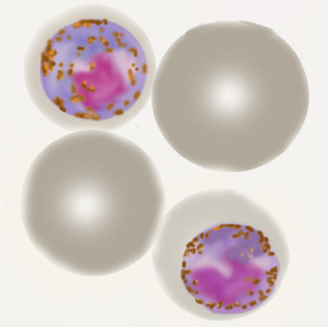
| Plasmodium knowlesi |
Early trophozoite
Late trophozoite
Schizont

Gametocyte
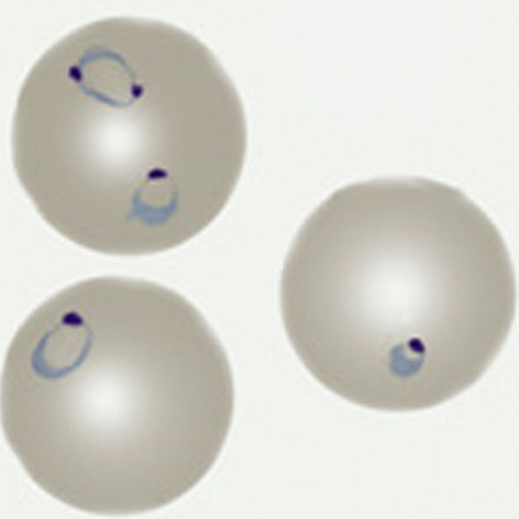

Brief Summary
- Very limited geographical distribution within S.E Asia
- Small fine ring forms resemble those of P.falciparum and may have high parasite count
- Later rings are more solid or elongated similar to P.malariae, although faint dots may be present
- Schizonts & gametocytes are often present and may resemble P.malariae but are less "neat"
- Characteristically red cell size is unaffected, although distortion may be seen
For more information
- click for full description of P.knowlesis morphology
- haga clic para obtener una descripción completa de la morfología de P.knowlesis
- click to visit the gallery of P.knowlesi forms