Schizont Development: Difference between revisions
From haematologyetc.co.uk
No edit summary |
No edit summary |
||
| Line 24: | Line 24: | ||
</gallery> | </gallery> | ||
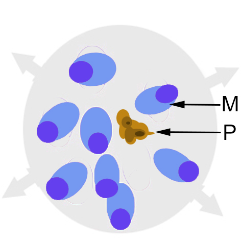
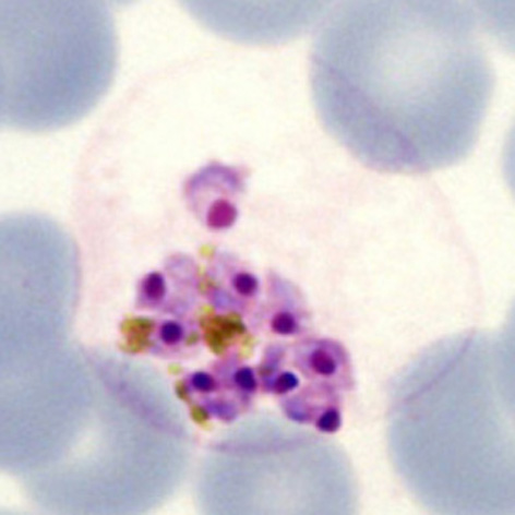
The cartoon image (A) shows the division of chromatin into two masses within a continuous blue parasite cytoplams (indiviual merozoites are not really distinguishable here). A clinical image of a parasite at this developmental stage (''P.ovale'' with well shown James'dots) is shown in panel (B). | |||
Revision as of 12:00, 27 March 2024
Navigation
Go Back
| How does schizont appearance change during their development?
THE INITIAL ASEXUAL DIVISION
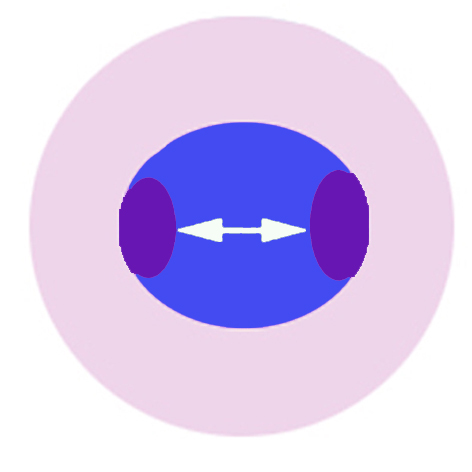
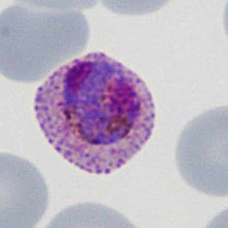
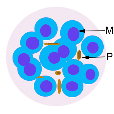
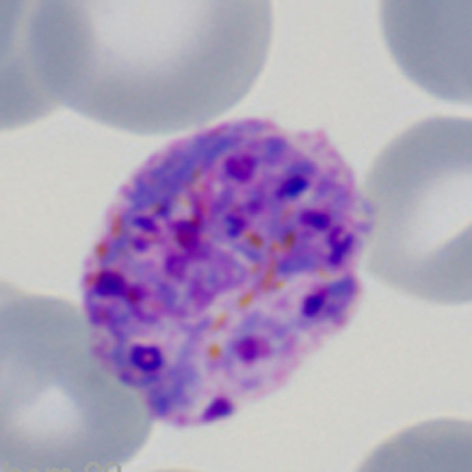
The cartoon image (A) shows the division of chromatin into two masses within a continuous blue parasite cytoplams (indiviual merozoites are not really distinguishable here). A clinical image of a parasite at this developmental stage (P.ovale with well shown James'dots) is shown in panel (B).
IMMATURE SCHIZONT APPEARANCES
xxxxxx.
MATURE SCHIZONT APPEARANCES xxxxxx
xxxxxx.
xxxxxx
xxxxxx. |