Schizont Development: Difference between revisions
From haematologyetc.co.uk
No edit summary |
No edit summary |
||
| (5 intermediate revisions by the same user not shown) | |||
| Line 9: | Line 9: | ||
The schizont form we see reflects successive cycles of asexual division that eventually result in the formation of multiple separate "merozoite" forms that are | The schizont form we see reflects successive cycles of asexual division that eventually result in the formation of multiple separate "merozoite" forms that are released as the red cell breaks down. Each merozoite then goes on to infect another red cell. This is therefore a progressive process and schizonts will look very different depending on which stage of development they represent. Below are images of schizonts at different developmental stages. | ||
---- | ---- | ||
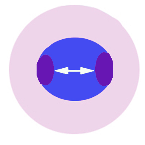
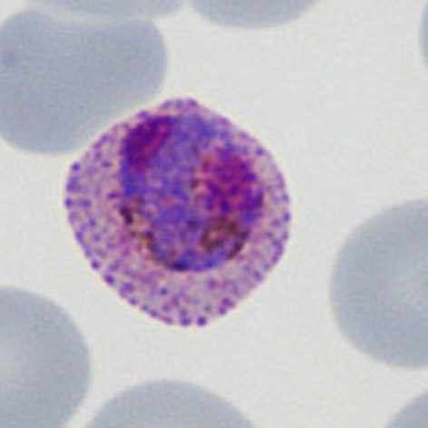
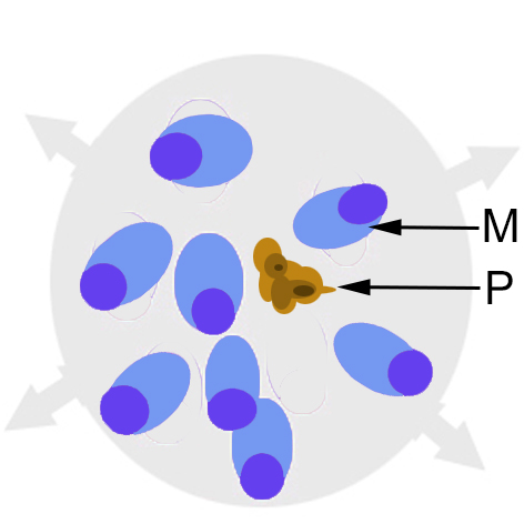
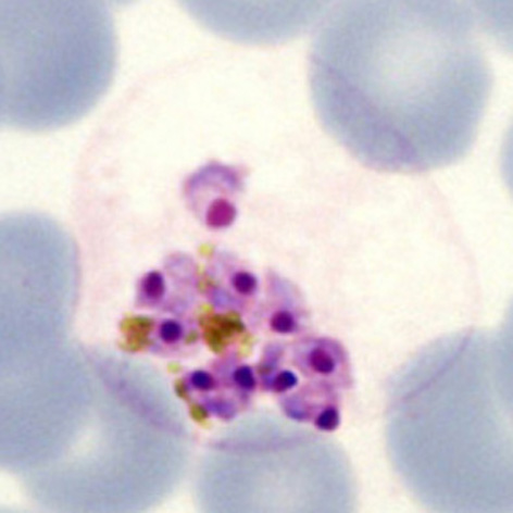
''' | '''THE INITIAL ASEXUAL DIVISION''' | ||
xxxxxx | |||
<gallery mode="nolines" widths="200px" heights="220px" > | <gallery mode="nolines" widths="200px" heights="220px" > | ||
File: | File:Schizontcartoon1.jpg|A|link={{filepath:Schizontcartoon1.jpg}} | ||
File: | File:Schizontreal1.jpg|B|link={{filepath:Schizontreal1.jpg}} | ||
</gallery> | </gallery> | ||
xxxxxx. | |||
---- | ---- | ||
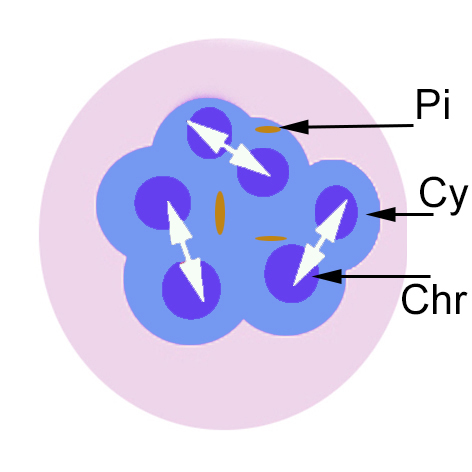
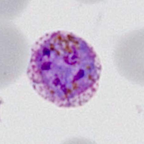
''' | '''IMMATURE SCHIZONT APPEARANCES''' | ||
xxxxxx | |||
<gallery mode="nolines" widths="200px" heights="220px" > | <gallery mode="nolines" widths="200px" heights="220px" > | ||
File: | File:Schizontcartoon2.jpg|A|link={{filepath:Schizontcartoon2.jpg}} | ||
File: | File:Schizontreal2.jpg|B|link={{filepath:Schizontreal2.jpg}} | ||
</gallery> | </gallery> | ||
xxxxxx. | |||
---- | ---- | ||
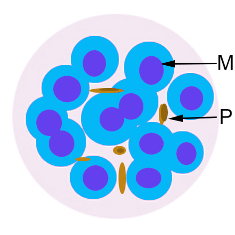
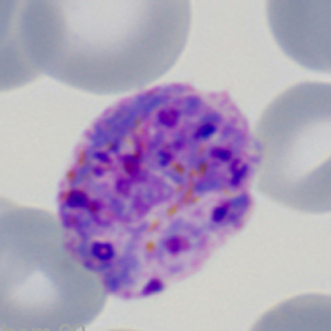
''' | '''MATURE SCHIZONT APPEARANCES''' | ||
xxxxxx | |||
<gallery mode="nolines" widths="200px" heights="220px" > | <gallery mode="nolines" widths="200px" heights="220px" > | ||
File: | File:Schizontcartoon3.jpg|A|link={{filepath:Schizontcartoon3.jpg}} | ||
File: | File:Schizontreal3.jpg|B|link={{filepath:Schizontreal3.jpg}} | ||
</gallery> | </gallery> | ||
xxxxxx. | |||
---- | ---- | ||
''' | '''MEROZOITE RELEASE''' | ||
xxxxxx | |||
<gallery mode="nolines" widths="200px" heights="220px" > | <gallery mode="nolines" widths="200px" heights="220px" > | ||
File: | File:Schizontcartoon4.jpg|A|link={{filepath:Schizontcartoon4.jpg}} | ||
File: | File:Schizontreal4.jpg|B|link={{filepath:Schizontreal4.jpg}} | ||
</gallery> | </gallery> | ||
xxxxxx. | |||
Revision as of 13:27, 26 March 2024
Navigation
Go Back
| How does schizont appearance change during their development?
THE INITIAL ASEXUAL DIVISION
xxxxxx.
IMMATURE SCHIZONT APPEARANCES
xxxxxx.
MATURE SCHIZONT APPEARANCES xxxxxx
xxxxxx.
xxxxxx
xxxxxx. |