Pre-erythrocytic (liver) stage: Difference between revisions
From haematologyetc.co.uk
No edit summary |
No edit summary |
||
| (11 intermediate revisions by the same user not shown) | |||
| Line 9: | Line 9: | ||
{| class="wikitable" style="border-style: solid; border-width: 5px; border-color: #023020; color:black" | {| class="wikitable" style="border-style: solid; border-width: 5px; border-color: #023020; color:black" | ||
|colspan="1" style = "font-size:100%; color:black; background: #afbddb |'''The | |colspan="1" style = "font-size:100%; color:black; background: #afbddb |'''The initial infection of the host''' | ||
|} | |} | ||
<gallery mode="nolines" widths=300px heights=300px> | <gallery mode="nolines" widths=300px heights=300px> | ||
File:Liver 0.jpg|<span style="font-size: | File:Liver 0.jpg|<span style="font-size:90%">''Infection of the mosquito''</span>|link={{filepath:Liver 0.jpg}} | ||
</gallery> | </gallery> | ||
The initial mosquito bite (Image A): blood from the host is taken through as mosquito mouthparts (m) from small vessels (v). At the same time fluid from mosquito salivary glands passes into the vessel. If a mosquitos is host to the malaria organism then parasites enter the blood with this saliva (in the form of '''"sporozoites"''' (sp)). | |||
---- | ---- | ||
{| class="wikitable" style="border-style: solid; border-width: 5px; border-color: #023020; color:black" | {| class="wikitable" style="border-style: solid; border-width: 5px; border-color: #023020; color:black" | ||
|colspan="1" style = "font-size:100%; color:black; background: #afbddb |''' | |colspan="1" style = "font-size:100%; color:black; background: #afbddb |'''Infection and replication in liver''' | ||
|} | |} | ||
<gallery mode="nolines" widths=300px heights=300px> | <gallery mode="nolines" widths=300px heights=300px> | ||
File: | File:Liver 1.jpg|<span style="font-size:90%">''Formation and release of merozoites in liver''</span>|link={{filepath:Liver 1.jpg}} | ||
</gallery> | </gallery> | ||
The sporozoites that have entered the blood then pass through the blood vessel and into the cells of the liver (B). They may pass through multiple cells but then remain in a single liver cell (C). | |||
---- | ---- | ||
{| class="wikitable" style="border-style: solid; border-width: 5px; border-color: #023020; color:black" | {| class="wikitable" style="border-style: solid; border-width: 5px; border-color: #023020; color:black" | ||
|colspan="1" style = "font-size:100%; color:black; background: #afbddb |''' | |colspan="1" style = "font-size:100%; color:black; background: #afbddb |'''From liver to blood''' | ||
|} | |} | ||
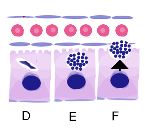
The parasites in liver cells (D) then undergo repeated cycles of asexual replication (E) to form schizonts which are similar to the schizont stage in blood. At the end of the process the '''“merozoites”''' that are formed are released into blood (F). | |||
<gallery mode="nolines" widths=" | <gallery mode="nolines" widths="300px" heights="300px" > | ||
File:Liver 2.jpg|Mature schizont releasing merozoites|link={{filepath:Liver 2.jpg}} | File:Liver 2.jpg|<span style="font-size:90%">''Mature schizont releasing merozoites''|link={{filepath:Liver 2.jpg}} | ||
</gallery> | </gallery> | ||
<gallery mode="nolines" widths="200px" heights=" | |||
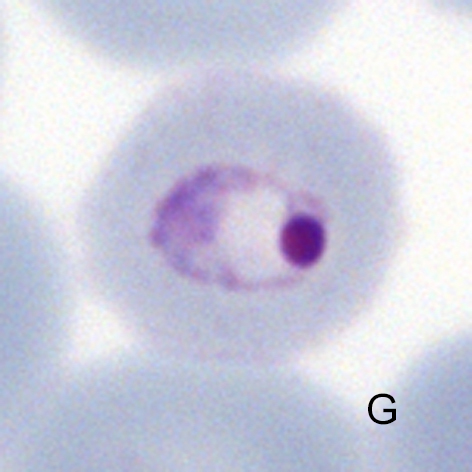
File:Liver 3.jpg| | They then infect red cells and now have the typical appearance of early trophozoite (G). | ||
<gallery mode="nolines" widths="200px" heights="200px" > | |||
File:Liver 3.jpg|<span style="font-size:90%">''Trophozoites now appear in blood''|link={{filepath:Liver 3.jpg}} | |||
</gallery> | </gallery> | ||
| Line 47: | Line 57: | ||
{| class="wikitable" style="border-style: solid; border-width: 5px; border-color: #023020; color:black" | {| class="wikitable" style="border-style: solid; border-width: 5px; border-color: #023020; color:black" | ||
|colspan="1" style = "font-size:100%; color:black; background: #afbddb |'''Relevance of | |colspan="1" style = "font-size:100%; color:black; background: #afbddb |'''Relevance of hepatic stage to clinical biology''' | ||
|} | |} | ||
The hepatic stage is the period where parasites begin to replicate and cause infection - this corresponds to an incubation period before symptoms begin; this will typically last between 1-4 weeks, and during this time parasites will not be detected in blood. The number of bites required for infection may be only 1 if the mosquito is heavily infected (hence cases of airport malaria where the infection is acquired from a mosquito passenger in luggage). | |||
For two malaria species (''P.ovale'' and ''P.vivax'') the hepatic stage may lie dormant for a period of time - this is the hynozoite (“sleeping animal”). This hyponozoite may reactivate - typically in less than a year although occasionally longer, causing clinical malaria symptoms long after the initial infection. | |||
---- | ---- | ||
Latest revision as of 11:01, 22 April 2024
Navigation
(click blue highlighted text to return to page)
Malaria main index
>Basic malaria biology
>>This page: Pre-erythrocytic (liver) stage
| The initial infection of the host |
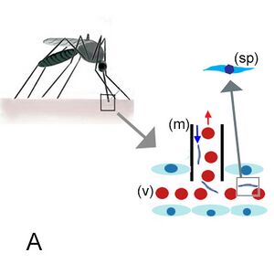
Infection of the mosquito
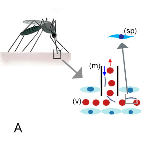
The initial mosquito bite (Image A): blood from the host is taken through as mosquito mouthparts (m) from small vessels (v). At the same time fluid from mosquito salivary glands passes into the vessel. If a mosquitos is host to the malaria organism then parasites enter the blood with this saliva (in the form of "sporozoites" (sp)).
| Infection and replication in liver |
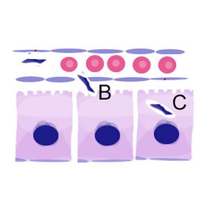
Formation and release of merozoites in liver

The sporozoites that have entered the blood then pass through the blood vessel and into the cells of the liver (B). They may pass through multiple cells but then remain in a single liver cell (C).
| From liver to blood |
The parasites in liver cells (D) then undergo repeated cycles of asexual replication (E) to form schizonts which are similar to the schizont stage in blood. At the end of the process the “merozoites” that are formed are released into blood (F).

Mature schizont releasing merozoites
They then infect red cells and now have the typical appearance of early trophozoite (G).
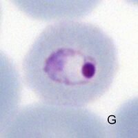
Trophozoites now appear in blood
| Relevance of hepatic stage to clinical biology |
The hepatic stage is the period where parasites begin to replicate and cause infection - this corresponds to an incubation period before symptoms begin; this will typically last between 1-4 weeks, and during this time parasites will not be detected in blood. The number of bites required for infection may be only 1 if the mosquito is heavily infected (hence cases of airport malaria where the infection is acquired from a mosquito passenger in luggage).
For two malaria species (P.ovale and P.vivax) the hepatic stage may lie dormant for a period of time - this is the hynozoite (“sleeping animal”). This hyponozoite may reactivate - typically in less than a year although occasionally longer, causing clinical malaria symptoms long after the initial infection.