Plasmodium vivax: Morphology: Difference between revisions
From haematologyetc.co.uk
No edit summary |
No edit summary |
||
| (13 intermediate revisions by one other user not shown) | |||
| Line 42: | Line 42: | ||
<gallery mode="nolines" widths=200px heights=200px> | <gallery mode="nolines" widths=200px heights=200px> | ||
File: | File:PVLTc.jpg|link={{filepath:PVLTc.jpg}} | ||
File: | File:PVLT main.jpg|link={{filepath:PVLT main.jpg}} | ||
</gallery> | </gallery> | ||
<br clear=all> | <br clear=all> | ||
| Line 49: | Line 49: | ||
The later growth stage | The later growth stage during which parasites grow considerably and lose their ring appearance, this process is accompanied by substantial modification of the red cell and metabolism of it's haemoglobin to form malaria pigment. | ||
*infected erythrocytes become significantly enlarged and irregular in shape | *infected erythrocytes become significantly enlarged and irregular in shape | ||
*parasites lose their ring appearnace becoming irregular and "amoeboid" in form | *parasites lose their ring appearnace becoming irregular and "[[amoeboid]]" in form | ||
*numerous red/purple | *numerous red/purple Schüffner's dots are predent in the cytoplasm of red cells | ||
*malaria pigment is often present and has an irregular distribution | *[[malaria pigment]] is often present and has an irregular distribution | ||
<div style="width: 350px"> | <div style="width: 350px"> | ||
{| class="wikitable" style="border-left:solid 4px navy;border-right:solid 4px navy;border-top:solid 4px navy;border-bottom:solid 4px navy; font-size:90%; color:navy; align:center" | {| class="wikitable" style="border-left:solid 4px navy;border-right:solid 4px navy;border-top:solid 4px navy;border-bottom:solid 4px navy; font-size:90%; color:navy; align:center" | ||
| colspan="1"''|[[P. | | colspan="1"''|[[P.vivax late trophozoites gallery|Click for ''P.vivax'' late trophozoite gallery]]'' | ||
|} | |} | ||
</div> | </div> | ||
| Line 72: | Line 72: | ||
<gallery mode="nolines" widths=200px heights=200px> | <gallery mode="nolines" widths=200px heights=200px> | ||
File: | File:PVSc.jpg|link={{filepath:PFSc.jpg}} | ||
File: | File:PVS main.jpg|link={{filepath:PFS main.jpg}} | ||
</gallery> | </gallery> | ||
<br clear=all> | <br clear=all> | ||
The asexual form | The asexual stage of [[malaria parasite development]] - only some trophozoites form schizonts, but those that do undergo successive cycles of replication within the red cell to generate multiple [["merozoites"]] that then each invade a new red cell to continue and increase the infection. | ||
* | *a range of maturing schizonts will generally be present within enlarged red cells | ||
* | *when mature schizonts may contain 16-24 separate merozoites | ||
*[[ | *[[Schüffner's dots]] can be detected in any residual cytoplasm of the erythrocyte | ||
* | *[[Malaria pigment|malaria pigment]] is visible in irregularly distributed clumps over the schizont surface | ||
<div style="width: 350px"> | <div style="width: 350px"> | ||
{| class="wikitable" style="border-left:solid 4px navy;border-right:solid 4px navy;border-top:solid 4px navy;border-bottom:solid 4px navy; font-size:90%; color:navy; align:center" | {| class="wikitable" style="border-left:solid 4px navy;border-right:solid 4px navy;border-top:solid 4px navy;border-bottom:solid 4px navy; font-size:90%; color:navy; align:center" | ||
| colspan="1"''|[[P. | | colspan="1"''|[[P.vivax schizont gallery|Click for ''P.vivax'' schizont gallery]]'' | ||
|} | |} | ||
</div> | </div> | ||
| Line 103: | Line 102: | ||
<gallery mode="nolines" widths=200px heights=200px> | <gallery mode="nolines" widths=200px heights=200px> | ||
File: | File:PVGc.jpg|link={{filepath:PVGc.jpg}} | ||
File: | File:PVG main.jpg|link={{filepath:PVG main.jpg}} | ||
</gallery> | </gallery> | ||
<br clear=all> | <br clear=all> | ||
| Line 112: | Line 111: | ||
The sexual replication form (very distinctive). | The sexual replication form (very distinctive). | ||
* | *red cells are very large and have ovoid or distorted forms | ||
* | *macrogametocytes (female form) will often entirely fill the erythrocyte | ||
* | *microgametocytes (male form) have a cytoplasmic rim with visible Schüffner's dots | ||
* | *[[Malaria pigment|malaria pigment]] is clumped evenly over the surface of the gametocyte | ||
<div style="width: 350px"> | <div style="width: 350px"> | ||
{| class="wikitable" style="border-left:solid 4px navy;border-right:solid 4px navy;border-top:solid 4px navy;border-bottom:solid 4px navy; font-size:90%; color:navy; align:center" | {| class="wikitable" style="border-left:solid 4px navy;border-right:solid 4px navy;border-top:solid 4px navy;border-bottom:solid 4px navy; font-size:90%; color:navy; align:center" | ||
| colspan="1"''|[[P. | | colspan="1"''|[[P.vivax gametocyte gallery|Click for ''P.vivax'' gametocyte gallery]]'' | ||
|} | |} | ||
</div> | </div> | ||
| Line 127: | Line 125: | ||
---- | ---- | ||
Latest revision as of 14:17, 11 April 2024
Navigation
(click blue highlighted text to return to page)
Malaria main index
>Species identification: summary page
>>This page: P.vivax: morphology
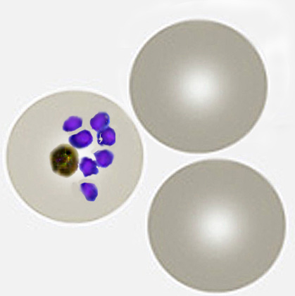
| The early trophozoite |
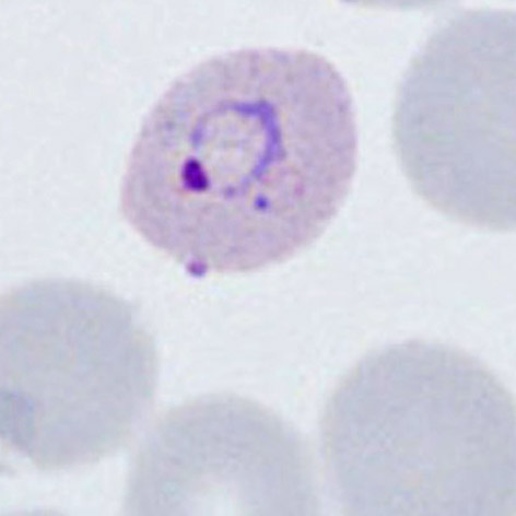
The earliest ring forms may be indistinguishable from other species, but during this stage the parasite tends to aquire a more irregular forms and to show signs of modification of the erythrocyte (added dots, and altered size and shape).
- erythrocytes begin to show increased size and altered shape
- parasites retain a ring form but may aquire a more irregular form
- parasites are generally large - occupying up to half of the erythrocyte
- cytoplasmic Schüffner's dots may appear at this stage, although pigment is less uncommon
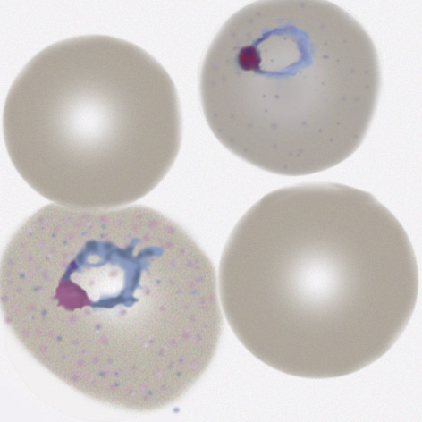
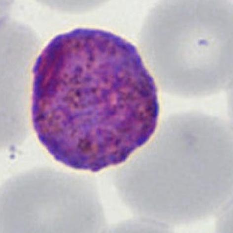
| The late trophozoite |
The later growth stage during which parasites grow considerably and lose their ring appearance, this process is accompanied by substantial modification of the red cell and metabolism of it's haemoglobin to form malaria pigment.
- infected erythrocytes become significantly enlarged and irregular in shape
- parasites lose their ring appearnace becoming irregular and "amoeboid" in form
- numerous red/purple Schüffner's dots are predent in the cytoplasm of red cells
- malaria pigment is often present and has an irregular distribution
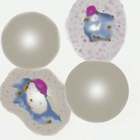
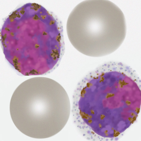
| The schizont |
The asexual stage of malaria parasite development - only some trophozoites form schizonts, but those that do undergo successive cycles of replication within the red cell to generate multiple "merozoites" that then each invade a new red cell to continue and increase the infection.
- a range of maturing schizonts will generally be present within enlarged red cells
- when mature schizonts may contain 16-24 separate merozoites
- Schüffner's dots can be detected in any residual cytoplasm of the erythrocyte
- malaria pigment is visible in irregularly distributed clumps over the schizont surface
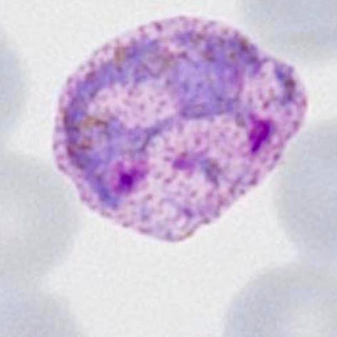
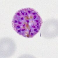
The gametocyte
| The gametocyte |
The sexual replication form (very distinctive).
- red cells are very large and have ovoid or distorted forms
- macrogametocytes (female form) will often entirely fill the erythrocyte
- microgametocytes (male form) have a cytoplasmic rim with visible Schüffner's dots
- malaria pigment is clumped evenly over the surface of the gametocyte