Plasmodium falciparum: Morphology: Difference between revisions
From haematologyetc.co.uk
No edit summary |
No edit summary |
||
| Line 2: | Line 2: | ||
'''Navigation'''</br> | '''Navigation'''</br> | ||
<span style="font-size:90%">[[Malaria Index|Malaria main index]] | <span style="font-size:90%">[[Malaria Index|Malaria main index]]</span></br> | ||
<span style="font-size:90%">[[Species Indentification: summary page | <span style="font-size:90%">[[Species Indentification: summary page]]</span></br> | ||
<span style="font-size:90%">''Plasmodium falciparum'': morphology</span> | <span style="font-size:90%">>''Plasmodium falciparum'': morphology</span> | ||
---- | ---- | ||
Revision as of 11:33, 18 March 2024
Navigation
Malaria main index
Species Indentification: summary page
>Plasmodium falciparum: morphology
Navigation
- Malaria main index
- Species identification: summary page
- Plasmodium falciparum: morphology
- Species identification: summary page
Geographical distribution
P.falciparum infection occurs in tropical and subtropical areas of central and South America, Africa, and S.E.Asia; this resembles the distribution of P.malariae and overlaps but is distinct from the distribution of P.vivax and P.ovale.
Detailed geographical information may be accessed here: [1].
The early trophozoite
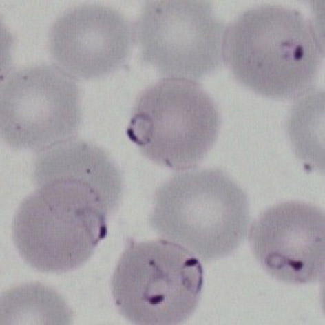
The earliest developing stage, and often the only form present in this species:
- Ring forms that are fine and delicate
- Frequently the red cells contain multiple parasites
- Parasites may have a distinctive double chromatin dot (signet ring form)
- Parasites may appear on the edge of the red cell and have a flattened appearance (accolé forms)
- Affected red cells have normal size and haemoglobin content
The late trophozoite
rleft
The later developing stage:
- Parasites resemble early ring forms, but are thicker and slightly larger
- Additional dots and clefts in cytoplasm when stained correctly (blue and relatively low in number).
- These have a characteristic appearance and are called Maurer's dots and clefts
- Size and shape of infected red cells usually unaffected, but may become crenated
- Look for double chromatin dot, Accolé forms, multiple parasites/cell
The schizont
The asexual replication stage:
- Do not generally circulate in this species unless overwhelming infection
- Contain multiple asexually formed developing parasites (most frequently 8-16)
- Development is progressive: first there are multiple chromatin dots, later a distinct nucleus and cytoplasm appears
- Loose pigment may be seen in clumps between the parasites
- Red cell size is generally unaffected but haemoglobin will largely be absent (metabolised by the parasites)
The gametocyte
The sexual replication stage (very distinctive).
- Gametocytes are elongated but are also restricted by the red cell membrane
- They appear as straight rods but frequently curve into a “banana form”
- The residual membrane (empty of haemoglobin) may appear as a "blister" to the side of the parasite
- The single chromatin area is in the centre of the parasite, often pigment overlies or surrounds it
- Gametocytes may not be seen in many cases.
Gallery
Click here to see gallery of Plasmodium falciparum forms