P.vivax late trophozoites gallery: Difference between revisions
From haematologyetc.co.uk
(Created page with "---- '''Navigation'''</br> Go Back ---- {| class="wikitable" style="border-style: solid; border-width: 4px; color:black" |colspan="1" style = "font-size:100%; color:black; background: FFFAFA"|<span style="color:black> {| class="wikitable" style="border-style: solid; border-width: 0px; border-color: #023020; color:black" |colspan="1" style = "font-size:100%; color:black; background: CBD5CO |'''''P.vivax'' gallery of early trophozoites''''...") |
No edit summary |
||
| (3 intermediate revisions by the same user not shown) | |||
| Line 7: | Line 7: | ||
|colspan="1" style = "font-size:100%; color:black; background: FFFAFA"|<span style="color:black> | |colspan="1" style = "font-size:100%; color:black; background: FFFAFA"|<span style="color:black> | ||
{| class="wikitable" style="border-style: solid; border-width: 0px; border-color: #023020; color:black" | {| class="wikitable" style="border-style: solid; border-width: 0px; border-color: #023020; color:black" | ||
|colspan="1" style = "font-size:100%; color:black; background: CBD5CO |'''''P.vivax'' gallery of | |colspan="1" style = "font-size:100%; color:black; background: CBD5CO |'''''P.vivax'' gallery of late trophozoites''''' | ||
|} | |} | ||
<span style="font-size:95%">'''Summary'''</span> | <span style="font-size:95%">'''Summary'''</span> | ||
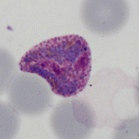
<span style="font-size:95%"> | <span style="font-size:95%">There is no absolutely clear point at which early trophozoites of ''P.vivax'' become late trophozoites as it is simply a process of continuous development. However, the transition to an amoeboid parasite form where the original ring structure can no longer be seen will certainly place the parasites at this stage. | ||
---- | ---- | ||
<gallery mode="traditional" widths=240px heights=240px> | <gallery mode="traditional" widths=240px heights=240px> | ||
File:PVLT1.jpg|<span style="font-size:80%">''' | File:PVLT1.jpg|<span style="font-size:80%">'''Developing late trophozoite''' The ring form is lost, the parasite is very irregular</span>|link={{filepath:PVLT1.jpg}} | ||
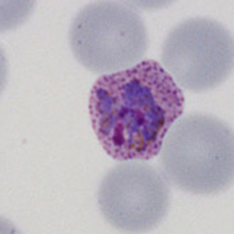
File:PVLT2.jpg|<span style="font-size:80%">''' | File:PVLT2.jpg|<span style="font-size:80%">'''Late trophozoite''' Note the solid parasite and the red cell begins to show distortion.</span>|link={{filepath:PVLT2.jpg}} | ||
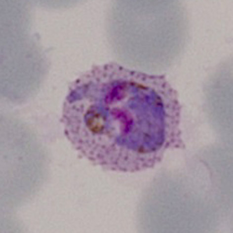
File:PVLT3.jpg|<span style="font-size:80%">''' | File:PVLT3.jpg|<span style="font-size:80%">'''Late trophozoite''' Similar to the previous example, the ring structure is entirely lost </span>|link={{filepath:PVLT3.jpg}} | ||
File:PVLT4.jpg|<span style="font-size:80%">''' | File:PVLT4.jpg|<span style="font-size:80%">'''Classical amoeboid form''' note also the very enlarged distorted red cell</span>|link={{filepath:PVLT4p.jpg}} | ||
</gallery>" | </gallery>" | ||
Latest revision as of 11:10, 4 April 2024
Navigation
Go Back
|