SummaryAt the very earliest point of growth, the malarial parasites may be difficult to distinguish from other early trophozoites. However, in this species a range of maturing forms are generally present and later forms begin to aquire characteristic features of the species - the trophozoites become larger and acquire thisckened and more irregular features. At the same time the red cells begin to enlarge and become less regular with the appearance of typical ----
Navigation Go Back
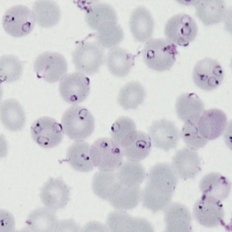
P.falciparum gallery of early trophozoites
SummaryAt this stage we look for typical (and often frequent) delicate rings within red cells that have normal (or slightly crenated) appearance. Forms often seen in this species include accolé forms, double chromatin dot forms, and multiple parasites within infected red cells.
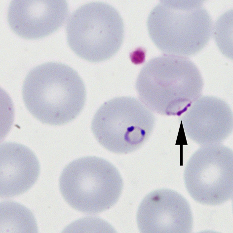
Fine ring form The small and delicate form of this species
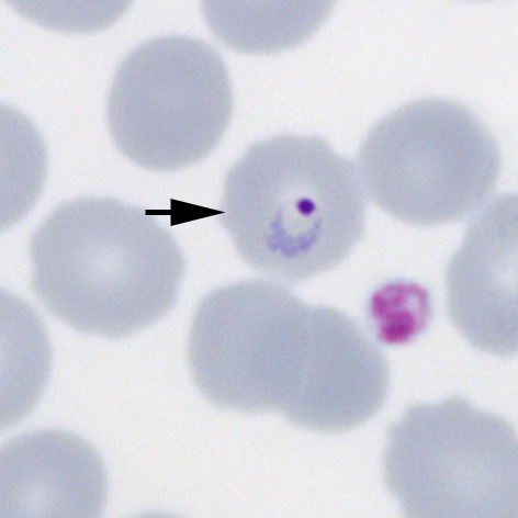
Double chromatin dot form Two chromatin dots (sometimes known as "signet ring" form).
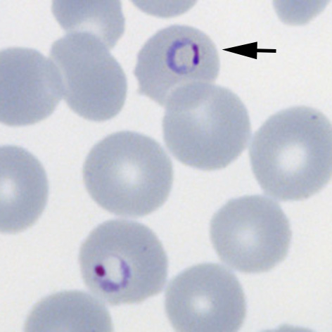
Accolé form: The arrowed form is closely associated with the red cell membrane
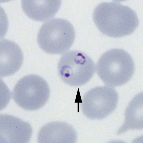
Multiple parasites Two parasites within a single red cells (arrowed)
High parasitaemia Most of the typical early trophozoite P.falciparum forms are present
"
Fine ring form The small and delicate form of this species
Double chromatin dot form Two chromatin dots (sometimes known as "signet ring" form).
Accolé form: The arrowed form is closely associated with the red cell membrane
Multiple parasites Two parasites within a single red cells (arrowed)
High parasitaemia Most of the typical early trophozoite P.falciparum forms are present