P.vivax early trophozoites gallery: Difference between revisions
From haematologyetc.co.uk
No edit summary |
No edit summary |
||
| Line 17: | Line 17: | ||
<gallery mode="traditional" widths=240px heights=240px> | <gallery mode="traditional" widths=240px heights=240px> | ||
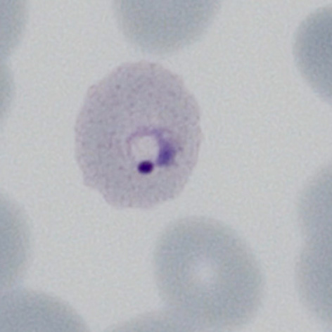
File:PVET1.jpg|<span style="font-size:80%">'''Very early | File:PVET1.jpg|<span style="font-size:80%">'''Very early form''' Very early red cell changes with fine dots visible</span>|link={{filepath:PVET1.jpg}} | ||
File:PVET2.jpg|<span style="font-size:80%">'''Early ring form''' Dots are more visible and the red cell begins to show distortion.</span>|link={{filepath:PVET2.jpg}} | File:PVET2.jpg|<span style="font-size:80%">'''Early ring form''' Dots are more visible and the red cell begins to show distortion.</span>|link={{filepath:PVET2.jpg}} | ||
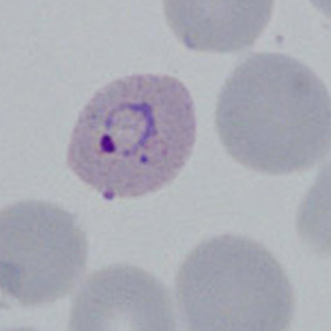
File:PVET3.jpg|<span style="font-size:80%">'''Early ring form''' The ring has thickened with established dots and altered red cell size</span>|link={{filepath:PVET3.jpg}} | File:PVET3.jpg|<span style="font-size:80%">'''Early ring form''' The ring has thickened with established dots and altered red cell size</span>|link={{filepath:PVET3.jpg}} | ||
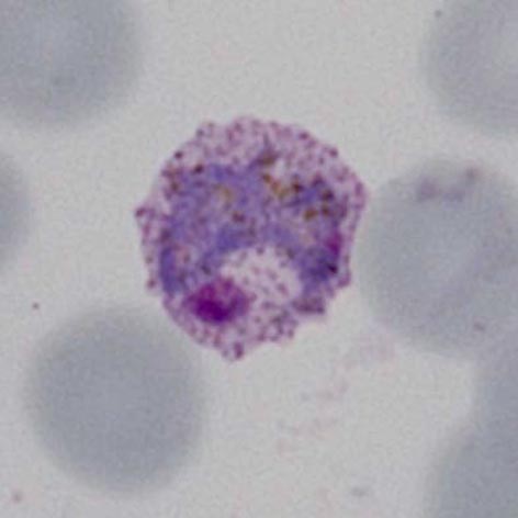
File:PVET4.jpg|<span style="font-size:80%">'''Intermediate ring''' | File:PVET4.jpg|<span style="font-size:80%">'''Intermediate ring''' Irregular parasite, possibly a double chromatin dot</span>|link={{filepath:PVET4p.jpg}} | ||
</gallery>" | </gallery>" | ||
Latest revision as of 11:12, 4 April 2024
Navigation
Go Back
|