P.vivax early trophozoites gallery: Difference between revisions
From haematologyetc.co.uk
(Created page with "---- '''Navigation'''</br> Go Back ---- {| class="wikitable" style="border-style: solid; border-width: 4px; color:black" |colspan="1" style = "font-size:100%; color:black; background: FFFAFA"|<span style="color:black> {| class="wikitable" style="border-style: solid; border-width: 0px; border-color: #023020; color:black" |colspan="1" style = "font-size:100%; color:black; background: CBD5CO |'''''P.vivax'' gallery of early trophozoites''''...") |
No edit summary |
||
| (11 intermediate revisions by the same user not shown) | |||
| Line 12: | Line 12: | ||
<span style="font-size:95%">'''Summary'''</span> | <span style="font-size:95%">'''Summary'''</span> | ||
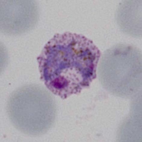
<span style="font-size:95%">At the very earliest point of growth, the malarial parasites may be difficult to distinguish from other early trophozoites. However, in this species a range of maturing forms are generally present and later forms begin to aquire characteristic features of the species - the trophozoites become larger and acquire thisckened and more irregular features. At the same time the red cells begin to enlarge and become less regular with the appearance of typical | <span style="font-size:95%">At the very earliest point of growth, the malarial parasites may be difficult to distinguish from other early trophozoites. However, in this species a range of maturing forms are generally present and later forms begin to aquire characteristic features of the species - the trophozoites become larger and acquire thisckened and more irregular features. At the same time the red cells begin to enlarge and become less regular with the appearance of typical Schuffner's dots in the red cell cytoplasm. | ||
' | |||
---- | ---- | ||
<gallery mode="traditional" widths=240px heights=240px> | <gallery mode="traditional" widths=240px heights=240px> | ||
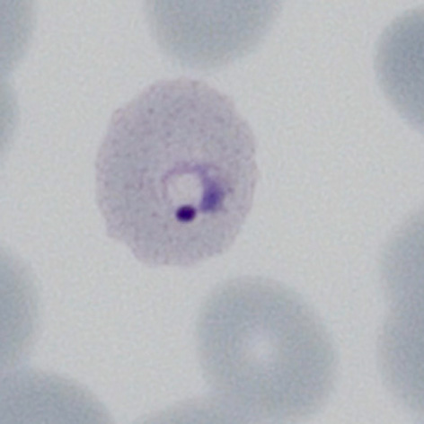
File: | File:PVET1.jpg|<span style="font-size:80%">'''Very early form''' Very early red cell changes with fine dots visible</span>|link={{filepath:PVET1.jpg}} | ||
File: | File:PVET2.jpg|<span style="font-size:80%">'''Early ring form''' Dots are more visible and the red cell begins to show distortion.</span>|link={{filepath:PVET2.jpg}} | ||
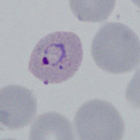
File: | File:PVET3.jpg|<span style="font-size:80%">'''Early ring form''' The ring has thickened with established dots and altered red cell size</span>|link={{filepath:PVET3.jpg}} | ||
File: | File:PVET4.jpg|<span style="font-size:80%">'''Intermediate ring''' Irregular parasite, possibly a double chromatin dot</span>|link={{filepath:PVET4p.jpg}} | ||
</gallery>" | </gallery>" | ||
Revision as of 11:12, 4 April 2024
Navigation
Go Back
|