P.falciparum schizont gallery: Difference between revisions
From haematologyetc.co.uk
(Created page with "---- '''Navigation'''</br> Go Back ---- {| class="wikitable" style="border-style: solid; border-width: 4px; color:black" |colspan="1" style = "font-size:100%; color:black; background: FFFAFA"|<span style="color:black> {| class="wikitable" style="border-style: solid; border-width: 0px; border-color: #023020; color:black" |colspan="1" style = "font-size:100%; color:black; background: CBD5CO |'''''P.falciparum'' gallery of late trophoz...") |
No edit summary |
||
| Line 18: | Line 18: | ||
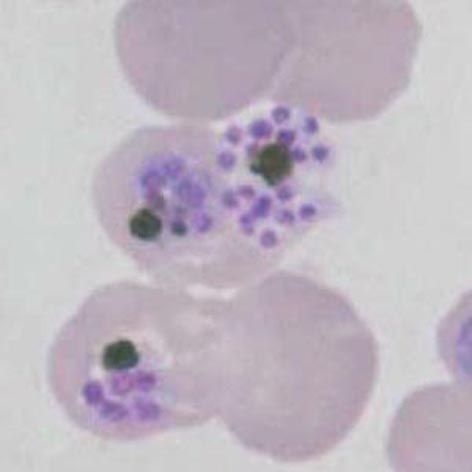
File:PFS1p.jpg|<span style="font-size:80%">'''Late rings''' Two cells both with typical dots: multiply infected and double dot forms</span>|link={{filepath:PFS1p.jpg}} | File:PFS1p.jpg|<span style="font-size:80%">'''Late rings''' Two cells both with typical dots: multiply infected and double dot forms</span>|link={{filepath:PFS1p.jpg}} | ||
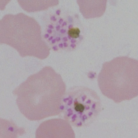
File:PFS2p.jpg|<span style="font-size:80%">'''Double chromatin dot form''' also Maurers dost and clefts, slight crenation and lost pallor</span>|link={{filepath:PFS2p.jpg}} | File:PFS2p.jpg|<span style="font-size:80%">'''Double chromatin dot form''' also Maurers dost and clefts, slight crenation and lost pallor</span>|link={{filepath:PFS2p.jpg}} | ||
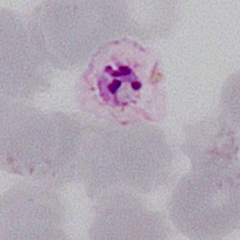
File: | File:PFS3p.jpg|<span style="font-size:80%">'''Accolé form''': closely associated with the red cell membrane, scanty mauers dots</span>|link={{filepath:PFS3p.jpg}} | ||
</gallery>" | </gallery>" | ||
Revision as of 00:42, 21 March 2024
Navigation
Go Back
|