P.falciparum late trophozoites gallery: Difference between revisions
From haematologyetc.co.uk
No edit summary |
No edit summary |
||
| Line 19: | Line 19: | ||
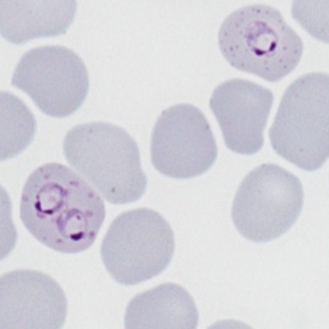
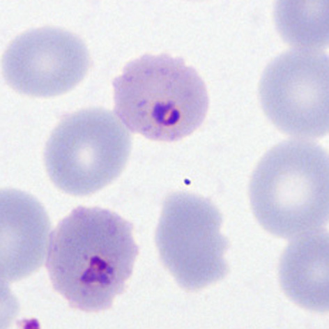
File:PFLT2p.jpg|<span style="font-size:80%">'''Double chromatin dot form''' also Maurers dost and clefts, slight crenation and lost pallor</span>|link={{filepath:PFLT2p.jpg}} | File:PFLT2p.jpg|<span style="font-size:80%">'''Double chromatin dot form''' also Maurers dost and clefts, slight crenation and lost pallor</span>|link={{filepath:PFLT2p.jpg}} | ||
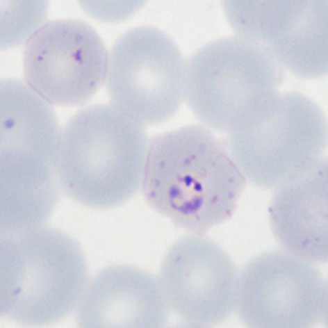
File:PFLT3p.jpg|<span style="font-size:80%">'''Accolé form''': closely associated with the red cell membrane, scanty mauers dots</span>|link={{filepath:PFLT3p.jpg}} | File:PFLT3p.jpg|<span style="font-size:80%">'''Accolé form''': closely associated with the red cell membrane, scanty mauers dots</span>|link={{filepath:PFLT3p.jpg}} | ||
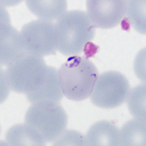
File:PFLT4p.jpg|<span style="font-size:80%">''' | File:PFLT4p.jpg|<span style="font-size:80%">'''Accolé form''' A nice typical form with scanty well-formed Maurers dots</span>|link={{filepath:PFLT4p.jpg}} | ||
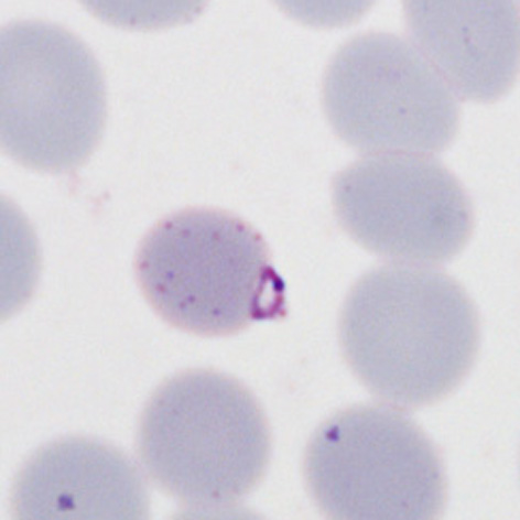
File:PFLT5p.jpg|<span style="font-size:80%">'''High parasitaemia''' Most of the typical early trophozoite ''P.falciparum'' forms are present</span>|link={{filepath:PFLT5p.jpg}} | File:PFLT5p.jpg|<span style="font-size:80%">'''High parasitaemia''' Most of the typical early trophozoite ''P.falciparum'' forms are present</span>|link={{filepath:PFLT5p.jpg}} | ||
</gallery>" | </gallery>" | ||
Revision as of 00:29, 21 March 2024
Navigation
Go Back
|