P.falciparum late trophozoites gallery: Difference between revisions
From haematologyetc.co.uk
No edit summary |
No edit summary |
||
| Line 16: | Line 16: | ||
---- | ---- | ||
<gallery mode="traditional" widths=240px heights=240px> | <gallery mode="traditional" widths=240px heights=240px> | ||
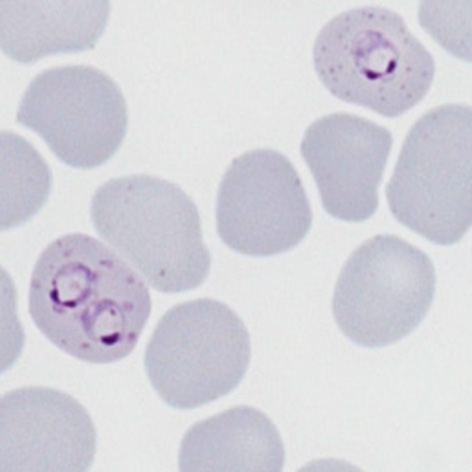
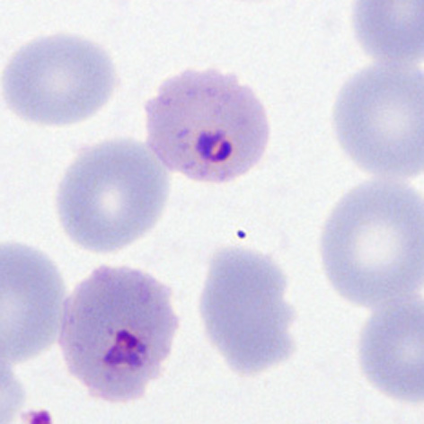
File:PFLT1p.jpg|<span style="font-size:80%">''' | File:PFLT1p.jpg|<span style="font-size:80%">'''Late rings''' Two cells both with typical dots: multiply infected and double dot forms</span>|link={{filepath:PFLT1p.jpg}} | ||
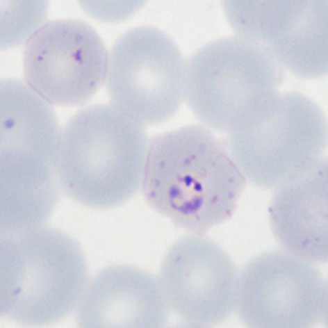
File:PFLT2p.jpg|<span style="font-size:80%">'''Double chromatin dot form''' Two chromatin dots (sometimes known as "signet ring" form).</span>|link={{filepath:PFLT2p.jpg}} | File:PFLT2p.jpg|<span style="font-size:80%">'''Double chromatin dot form''' Two chromatin dots (sometimes known as "signet ring" form).</span>|link={{filepath:PFLT2p.jpg}} | ||
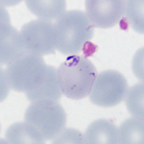
File:PFLT3p.jpg|<span style="font-size:80%">'''Accolé form''': The arrowed form is closely associated with the red cell membrane</span>|link={{filepath:PFLT3p.jpg}} | File:PFLT3p.jpg|<span style="font-size:80%">'''Accolé form''': The arrowed form is closely associated with the red cell membrane</span>|link={{filepath:PFLT3p.jpg}} | ||
Revision as of 00:24, 21 March 2024
Navigation
Go Back
|