P.falciparum gametocyte gallery: Difference between revisions
From haematologyetc.co.uk
No edit summary |
No edit summary |
||
| Line 7: | Line 7: | ||
|colspan="1" style = "font-size:100%; color:black; background: FFFAFA"|<span style="color:black> | |colspan="1" style = "font-size:100%; color:black; background: FFFAFA"|<span style="color:black> | ||
{| class="wikitable" style="border-style: solid; border-width: 0px; border-color: #023020; color:black" | {| class="wikitable" style="border-style: solid; border-width: 0px; border-color: #023020; color:black" | ||
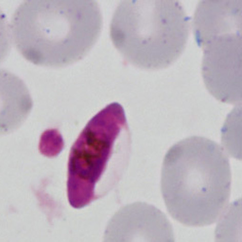
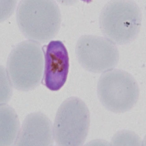
|colspan="1" style = "font-size:100%; color:black; background: CBD5CO |'''''P.falciparum'' gallery of | |colspan="1" style = "font-size:100%; color:black; background: CBD5CO |'''''P.falciparum'' gallery of gametocytes''''' | ||
|} | |} | ||
<span style="font-size:95%">'''Summary'''</span> | <span style="font-size:95%">'''Summary'''</span> | ||
<span style="font-size:95%"> | <span style="font-size:95%">Gametocytes in this species are highly distinctive. They develop within the red cell as long rods, however the red cell membrane continues to restrict them - the haemoglobin is fully metabolised so is not visible, but the residual membrane of the red cell "ghost" can usually be seen to the side of the red cell. The gametocytes differ with large (macrogametocyte) forms that become curved because of the red cell membrane restricting thir shape (banana form); the smaller (microgametocytes) retain the rod shape. | ||
---- | ---- | ||
Revision as of 10:55, 21 March 2024
Navigation
Go Back
|