P.falciparum early trophozoites gallery: Difference between revisions
From haematologyetc.co.uk
No edit summary |
No edit summary |
||
| Line 5: | Line 5: | ||
{| class="wikitable" style="border-style: solid; border-width: 4px; color:black" | {| class="wikitable" style="border-style: solid; border-width: 4px; color:black" | ||
|colspan="1" style = "font-size:100%; color:black; background: FFFAFA"|<span style="color: | |colspan="1" style = "font-size:100%; color:black; background: FFFAFA"|<span style="color:black> | ||
{| class="wikitable" style="border-style: solid; border-width: 5px; border-color: #023020; color:black" | {| class="wikitable" style="border-style: solid; border-width: 5px; border-color: #023020; color:black" | ||
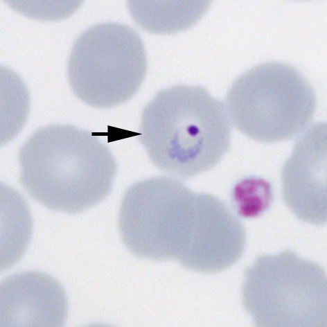
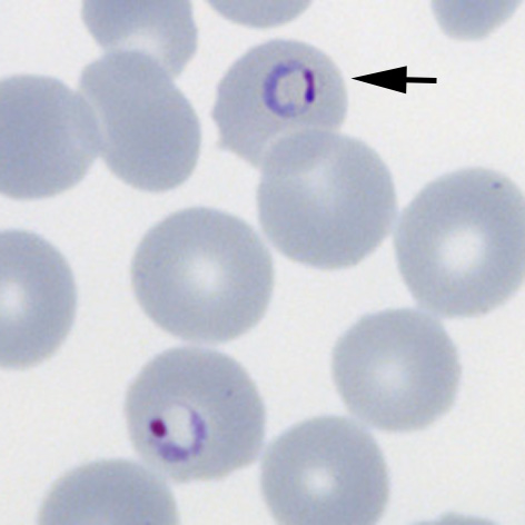
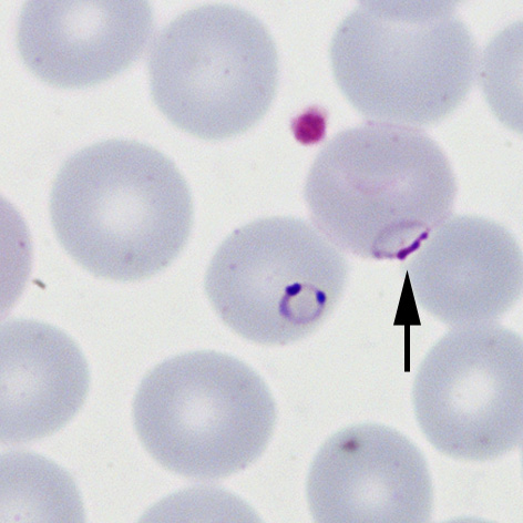
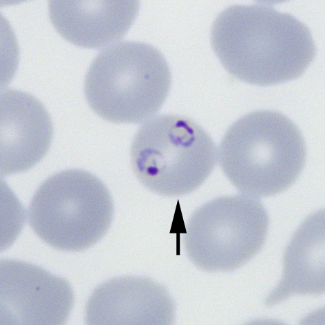
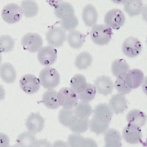
|colspan="1" style = "font-size:100%; color:black; background: CBD5CO |'''''P.falciparum'' gallery of early trophozoites''''' | |colspan="1" style = "font-size:100%; color:black; background: CBD5CO |'''''P.falciparum'' gallery of early trophozoites''''' | ||
Revision as of 12:52, 19 March 2024
Navigation
Go Back
|