MPAL and Acute leukaemia types: Difference between pages
From haematologyetc.co.uk
(Difference between pages)
No edit summary |
No edit summary |
||
| Line 1: | Line 1: | ||
---- | ---- | ||
<span style="font-size:110%; color:navy">'''SECTION 1: Establishing the primitive nature of the blast cells in AML'''</span> | |||
[[Image:AML M1.png|130px]] | |||
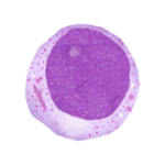
''Blast cells are recognised by morphology using features such as primitive nuclear or basophilic cytoplasmic appearances; similar principles are applied in flow cytometry where particular markers suggest a more primitive nature. | |||
<div style="width: 95%"> | |||
<div style="width: | {| class="mw-collapsible mw-collapsed wikitable" style="border-style: solid; border-width: 5px; color:black" </br>data-expandtext="Click to open Table" data-collapsetext="Click to close table" | ||
{| class="wikitable" style=" | ! colspan="2" style = "font-size:100%; color:black"| '''Table:''' Markers primarily used to confirm primitive nature in AML. | ||
!colspan="2" | |- | ||
|colspan="2" style = "font-size:90%; color:black; background:#ddeee1"|The expression of some markers is typically associated with primitive cells and can help recognise the primitive nature of blast cells.</br>Note that these markers may also have some lineage specificity but are not generally used to assign lineage | |||
|- | |- | ||
|colspan=" | |colspan="1" style = "font-size:90%; color:black;" |'''[[CD45]]''' | ||
|colspan="1" style = "font-size:90%;"|A marker expressed by all leukocytes and their precursors. In AML expression is characteristically "weak" i.e. significantly less intense than normal lymphocytes or monocytes. In monocytic AML expression may be stronger, particular in mare mature monocytic forms where expression may resemble normal monocytes. | |||
|- | |- | ||
|colspan="1" style = "font-size:90%; color:black;" |''' | |colspan="1" style = "font-size:90%; color:black;" |'''[[CD34]]''' | ||
|colspan="1" style = "font-size:90%;"| | |colspan="1" style = "font-size:90%;"|Frequently expressed by AML blast cells (40-80% of cases) - most often in less differentiated forms of AML. Expression is also seen frequently (and often more strongly) in ALL''' | ||
|- | |- | ||
|colspan="2" style = "font-size:90%; color:black; background: | |colspan="2" style = "font-size:90%; color:black; background:light gray" |'''Other markers:''' In the context of a proven AML diagnosis a number of markers may be expressed that reflect the primitive nature of the cells. Some of these are more frequently associated with lymphoid disorders, but providing other criteria for AML are met they should simply be considered to show "primitivness".</br>These include: [[CD38]], [[HLA-DR]], [[TDT]], [[CD7]]. | ||
|- | |- | ||
| | |} | ||
</div> | |||
---- | |||
<span style="font-size:110%; color:navy">'''SECTION 2: Assigning blast cells as myeloid lineage i.e. Diagnosing AML'''</span> | |||
[[Image:AML M2.png|150px]] | |||
''In morphology other features are used to indicate myeloid lineage, some are highly specificsuch as Auer rods, others may suggest myeloid lineage but be less specific. Again similar principles apply in flow cytometry'' | |||
There are two ways to make a diagnosis of AML | |||
<div style="width: 95%"; > | |||
{| class="wikitable" | |||
!colspan="2" style = "background:lightgrey; border:solid"| '''Requirements to diagnose AML by flow cytometry''' | |||
|- | |- | ||
!colspan="1" style = "background:#FFFFF0;border:solid; font-size:90%;"| '''Pattern 1:'''</br>A myeloid lineage-defining marker pattern is present</br>'''and'''</br>No lineage-defining markers of T or B cells are present | |||
!colspan="1" style = "background:white; border:solid; font-size:90%;"|See '''[[Table 1]]''' for lineage-defining marker patterns in AML | |||
|- | |||
!colspan="1" style = "background:#FFFFF0;border:solid; font-size:90%;"|'''Pattern 2''':</br>At least two myeloid lineage-associated markers are present</br>'''and'''</br>There are no lineage defining markers of T or B cells</br>'''and'''</br>No more than one: T-cell '''or''' B-cell lineage-associated markers are present | |||
!colspan="1" style = "background:white; border:solid; font-size:90%;"|See '''Table 2A''' and '''Table 2B''' for lineage-associated markers in AML | |||
|} | |} | ||
</div> | </div> | ||
<span style="font-size: | |||
<div style="width: 95%"> | |||
{| class="mw-collapsible mw-collapsed wikitable" style="border-style: solid; border-width: 5px; color:black" data-expandtext="Click to open Table" data-collapsetext="Click to close table" | |||
!colspan="2" <span style="font-size:100%; colour:white; text-align:left; border: 1px solid black; background:pale gray">|'''Table 1:''' Myeloid-lineage defining markers in AML </span><span style="font-size:100%; text-align:left; background:white"> | |||
|- | |||
* | |colspan="2" <span style = "font-size:90%; color:black; background:#ddeee1">|Lineage is defined by either the definite assignment to myeloid lineage or definite assignment to monocytic lineage. In each case there are specific criteria. | ||
</ | |- | ||
<span style="font-size: | |colspan="1" style = "font-size:90%; color:black;" |1. Definite myeloid linage marker: expression of '''[[MPO]]''' | ||
|colspan="1" style = "font-size:90%;"|A '''lineage-defining''' marker in AML when expressed (around 80% of cases). More frequent expressed in cases with granulocytic maturation. When detected by flow cytometry is diagnostic of myeloid differentiation is established. | |||
|- | |||
|colspan="1" style = "font-size:90%; color:black;" |2. Definite monocytic lineage, assigned if '''at least two''' of the following are expressed '''[[CD14]], [[CD11c]], [[CD64]], [[NSE]], lysozyme*''' | |||
|colspan="1" style = "font-size:90%;"|A '''two of these are lineage-defining''' in AML. Indicating monocytic maturation. *Note that NSE is a cytochemical stain. | |||
|} | |||
</div> | |||
<div style="width: 95%"> | |||
{| class="mw-collapsible mw-collapsed wikitable" style="border-style: solid; border-width: 5px; color:black" data-expandtext="Click to open Table" data-collapsetext="Click to close table" | |||
!colspan="2" <span style="font-size:100%; colour:white; text-align:left; border: 1px solid black; background:pale gray">|'''Table 2A:''' Myeloid lineage-associated markers (Main Set)'''</span><span style="font-size:90%; text-align:left; background:white"> | |||
|- | |||
|colspan="2" style = "font-size:90%; color:black; background:#ddeee1" |Each of these markers is frequently expressed in AML (80% of cases). They support a diagnosis of myeloid lineage, but are not sufficiently specific to be AML-defining when expressed alone.</br>These markers may be considered the most frequent markers expressed by AML, but are not alone sufficient to diagnose myeloid lineage.</br>However, in the absence of markers suggesting alternative lineage expression of '''two''' of these markers may be used to assign myeloid lineage. | |||
</br> | </br> | ||
|- | |||
|colspan="1" style = "font-size:90%; color:black;" |'''[[CD117]]''' | |||
|colspan="1" style = "font-size:84%;"|An early marker of myeloid lineage, seen in up to 80% of AML and vauable in recognising more primitive differentaiion forms (note that aberrant expression is seen in up to 20% of ALL cases) | |||
|- | |||
|colspan="1" style = "font-size:90%; color:black;" |'''[[CD33]]''' | |||
|colspan="1" style = "font-size:84%;"|A good marker for AML, particularly for those cases with granulocytic maturation, CD33 is often less strongly expressed in AML with monocytic dfferentiation and strongly expressed in APL. | |||
|- | |||
|colspan="1" style = "font-size:90%; color:black;" |'''[[CD13]]''' | |||
|colspan="1" style = "font-size:84%;"|A good lineage marker for AML that is acquired a little later in differentation than CD117 or CD33; expression of CD13 is often higher than CD33 in AML with monocytic differentiation. | |||
|- | |||
|} | |} | ||
</div> | </div> | ||
< | <div style="width: 95%"> | ||
{| class="mw-collapsible mw-collapsed wikitable" style="border-style: solid; border-width: 5px; color:black" data-expandtext="Click to open Table" data-collapsetext="Click to close table" | |||
!colspan="2" <span style="font-size:100%; colour:white; text-align:left; border: 1px solid black; background:pale gray">|'''Table 2B:''' Myeloid lineage-associated markers (Specific differentiation)'''</span><span style="font-size:90%; text-align:left; background:white"> | |||
---- | |- | ||
|colspan="2" style = "font-size:90%; color:black; background:#ddeee1" |The markers already described above are sufficient to make a diagnosis of typical AML in most cases. Typically the markers in this table are associated with specific features of differentiation so will not be present in all cases, but may be helpful as "lineage-assocated" markers in difficult cases. Care should be taken when doing so, since in some cases specificity may be lower than for typical myeloid markers. | |||
|- | |||
!colspan="2" style = "font-size:90%;background:white;"|Granulocytic lineage markers | |||
|- | |||
!colspan="2" | |colspan="1" style = "font-size:90%; color:black;" |'''[[CD11b]]''' | ||
|colspan="1" style = "font-size:84%;"|A marker of both granulocytic and monocytic maturation, this marker has previously been associated with less good outcome in a number of studies | |||
|- | |||
!colspan="2" style = "font-size:90%;background:white;"|Monocytic lineage markers | |||
|- | |||
|colspan="1" style = "font-size:90%; color:black;" |'''[[CD11c]]''' | |||
|colspan="1" style = "font-size:84%;"|This marker is most associated with monocytic maturation in AML being fairly well corellated with CD11B, but overall probably less specific for monocytic differentiation that CD14 | |||
|- | |||
|colspan="1" style = "font-size:90%; color:black;" |'''[[CD14]]''' | |||
|colspan="1" style = "font-size:84%;"|Primarily a marker of monocytic maturation in AML, seen most often in more differentiated forms, when present CD14 be considered a strong indicator of monocytic phenotype. | |||
|- | |||
|colspan="1" style = "font-size:90%; color:black;" |'''[[CD64]]''' | |||
|colspan="1" style = "font-size:84%;"|A good lineage marker for monocytic differentiation in AML, expressed in both monoblastic and monocytic forms, not fully sepicific when expressed at lower levels. | |||
|- | |||
!colspan="2" style = "font-size:90%;background:white;"|'''Features associated with erythroid differentiation''' | |||
|- | |||
|colspan="2" style = "font-size:90%;background:white;"|Most often [[CD34]], [[CD45]] and [[HLA-DR]] are weak or negative, although [[CD117]] and [[CD36]] are generally expressed. There may be some expression of platelet markers in some cases. | |||
|- | |- | ||
|colspan=" | |colspan="1" style = "font-size:90%; color:black;" |'''[[CD71]]''' | ||
|colspan="1" style = "font-size:84%;"|Frequently expressed though not fully lineage specific | |||
|- | |- | ||
|colspan="1" style = "font-size:90%; color:black;"| | |colspan="1" style = "font-size:90%; color:black;" |'''[[CD235]]''' | ||
|colspan="1" style = "font-size: | |colspan="1" style = "font-size:84%;"|A good marker of erythroid differentiation but acquired late and therefore may not be expressed | ||
|- | |- | ||
!colspan="2" style = "font-size:90%;background:white;"|'''Features associated with megakaryocytic differentiation''' | |||
|- | |- | ||
|colspan="2" style = "font-size:90%; | |colspan="2" style = "font-size:90%;background:white;"|Most often [[CD34]], [[CD45]] and [[HLA-DR]] are weak or negative, although [[CD13]] and [[CD33]] may be expressed | ||
|- | |- | ||
|colspan="1" style = "font-size:90%; color:black;"| | |colspan="1" style = "font-size:90%; color:black;" |'''[[CD41]]''' | ||
|colspan="1" style = "font-size: | |colspan="1" style = "font-size:84%;"|Platelet glycoprotein IIbIIIa | ||
|- | |- | ||
|colspan="1" style = "font-size:90%;"|''' | |colspan="1" style = "font-size:90%; color:black;" |'''[[CD61]]''' | ||
|colspan="1" style = "font-size: | |colspan="1" style = "font-size:84%;"|Platelet glycoprotein IIIa | ||
|- | |- | ||
|colspan="1" style = "font-size:90%; color:black;" |'''[[CD36]]''' | |||
|colspan="1" style = "font-size:84%;"|Relatively non-specific (seen in erythroid and monocytic leukaemias) but often strongly expressed | |||
</span> | |||
|} | |} | ||
</div> | </div> | ||
<span | |||
<div style="width: | ---- | ||
{| class="wikitable | |||
!colspan="2" | <span style="font-size:110%; color:navy">'''SECTION 3: When to consider alternative lineage assignment'''</span> | ||
The "aberrent" expression of lymphoid markers is frequently encountered in AML (and in some patterns of mutation these may be expected); however when non-myeloid markers are found it is important to consder if this changes lineage assignment. There are several conditions that should be considered: | |||
<div style="width: 95%"; font-size:90%> | |||
{| class="wikitable" | |||
!colspan="2" style = "background:lightgrey; border:solid"| '''When to consder an amended lineage assignment'''</br>The criteria below are brief and only indicate where concern should be raised, please see link (in blue) for full information. | |||
|- | |- | ||
|colspan=" | !colspan="1" style = "background:#FFFFF0;border:solid"| Mixed Phenotype Acute Leukaemia</br>Go to link</br>See: '''[[MPAL]]''' | ||
!colspan="1" style = "background:white;border:solid; font-size:90%;"| There is sufficient evidence to assign myeloid lineage '''but''' the cells also express either a lineage-defining marker of T or B cells, or more than one lineage associated marker of T or B cells. | |||
|- | |- | ||
!colspan="1" style = "background:#FFFFF0;border:solid"| Acute Undifferentiated Leukaenmia</br>See: '''[[AUL]]''' | |||
!colspan="1" style = "background:white; border:solid; font-size:90%;"| There is insufficient evidence to assign myeloid lineage, but there is expression of one myeloid-associated marker, '''and''' you cannot assign T-cell or B-cell lineage. I | |||
|- | |- | ||
!colspan="1" style = "background:#FFFFF0;border:solid"| Early T-cell precursor ALL</br>See: '''[[Non-haem]]''' | |||
!colspan="1" style = "background:white; border:solid; font-size:90%;"| Myeloid markers may occasionaly be expressed by non-haematological cells. Consider in atypical cases particularly where CD45 expression is very weak. | |||
|- | |- | ||
!colspan="1" style = "background:#FFFFF0;border:solid"| Non-haematological malignancy</br>See '''[[ETP-ALL]]''' | |||
!colspan="1" style = "background:white; border:solid; font-size:90%;"| The marker pattern is of a primitive T cell neoplasm with lineage-defining crieria, but there are also myeloid markers expressed | |||
|} | |} | ||
---- | |||
Revision as of 12:21, 22 November 2023
SECTION 1: Establishing the primitive nature of the blast cells in AML
Blast cells are recognised by morphology using features such as primitive nuclear or basophilic cytoplasmic appearances; similar principles are applied in flow cytometry where particular markers suggest a more primitive nature.
| Table: Markers primarily used to confirm primitive nature in AML. | |
|---|---|
| The expression of some markers is typically associated with primitive cells and can help recognise the primitive nature of blast cells. Note that these markers may also have some lineage specificity but are not generally used to assign lineage | |
| CD45 | A marker expressed by all leukocytes and their precursors. In AML expression is characteristically "weak" i.e. significantly less intense than normal lymphocytes or monocytes. In monocytic AML expression may be stronger, particular in mare mature monocytic forms where expression may resemble normal monocytes. |
| CD34 | Frequently expressed by AML blast cells (40-80% of cases) - most often in less differentiated forms of AML. Expression is also seen frequently (and often more strongly) in ALL |
| Other markers: In the context of a proven AML diagnosis a number of markers may be expressed that reflect the primitive nature of the cells. Some of these are more frequently associated with lymphoid disorders, but providing other criteria for AML are met they should simply be considered to show "primitivness". These include: CD38, HLA-DR, TDT, CD7. | |
SECTION 2: Assigning blast cells as myeloid lineage i.e. Diagnosing AML
In morphology other features are used to indicate myeloid lineage, some are highly specificsuch as Auer rods, others may suggest myeloid lineage but be less specific. Again similar principles apply in flow cytometry
There are two ways to make a diagnosis of AML
| Requirements to diagnose AML by flow cytometry | |
|---|---|
| Pattern 1: A myeloid lineage-defining marker pattern is present and No lineage-defining markers of T or B cells are present |
See Table 1 for lineage-defining marker patterns in AML |
| Pattern 2: At least two myeloid lineage-associated markers are present and There are no lineage defining markers of T or B cells and No more than one: T-cell or B-cell lineage-associated markers are present |
See Table 2A and Table 2B for lineage-associated markers in AML |
| Table 1: Myeloid-lineage defining markers in AML | |
|---|---|
| Lineage is defined by either the definite assignment to myeloid lineage or definite assignment to monocytic lineage. In each case there are specific criteria. | |
| 1. Definite myeloid linage marker: expression of MPO | A lineage-defining marker in AML when expressed (around 80% of cases). More frequent expressed in cases with granulocytic maturation. When detected by flow cytometry is diagnostic of myeloid differentiation is established. |
| 2. Definite monocytic lineage, assigned if at least two of the following are expressed CD14, CD11c, CD64, NSE, lysozyme* | A two of these are lineage-defining in AML. Indicating monocytic maturation. *Note that NSE is a cytochemical stain. |
| Table 2A: Myeloid lineage-associated markers (Main Set) | |
|---|---|
| Each of these markers is frequently expressed in AML (80% of cases). They support a diagnosis of myeloid lineage, but are not sufficiently specific to be AML-defining when expressed alone. These markers may be considered the most frequent markers expressed by AML, but are not alone sufficient to diagnose myeloid lineage. However, in the absence of markers suggesting alternative lineage expression of two of these markers may be used to assign myeloid lineage.
| |
| CD117 | An early marker of myeloid lineage, seen in up to 80% of AML and vauable in recognising more primitive differentaiion forms (note that aberrant expression is seen in up to 20% of ALL cases) |
| CD33 | A good marker for AML, particularly for those cases with granulocytic maturation, CD33 is often less strongly expressed in AML with monocytic dfferentiation and strongly expressed in APL. |
| CD13 | A good lineage marker for AML that is acquired a little later in differentation than CD117 or CD33; expression of CD13 is often higher than CD33 in AML with monocytic differentiation. |
| Table 2B: Myeloid lineage-associated markers (Specific differentiation) | |
|---|---|
| The markers already described above are sufficient to make a diagnosis of typical AML in most cases. Typically the markers in this table are associated with specific features of differentiation so will not be present in all cases, but may be helpful as "lineage-assocated" markers in difficult cases. Care should be taken when doing so, since in some cases specificity may be lower than for typical myeloid markers. | |
| Granulocytic lineage markers | |
| CD11b | A marker of both granulocytic and monocytic maturation, this marker has previously been associated with less good outcome in a number of studies |
| Monocytic lineage markers | |
| CD11c | This marker is most associated with monocytic maturation in AML being fairly well corellated with CD11B, but overall probably less specific for monocytic differentiation that CD14 |
| CD14 | Primarily a marker of monocytic maturation in AML, seen most often in more differentiated forms, when present CD14 be considered a strong indicator of monocytic phenotype. |
| CD64 | A good lineage marker for monocytic differentiation in AML, expressed in both monoblastic and monocytic forms, not fully sepicific when expressed at lower levels. |
| Features associated with erythroid differentiation | |
| Most often CD34, CD45 and HLA-DR are weak or negative, although CD117 and CD36 are generally expressed. There may be some expression of platelet markers in some cases. | |
| CD71 | Frequently expressed though not fully lineage specific |
| CD235 | A good marker of erythroid differentiation but acquired late and therefore may not be expressed |
| Features associated with megakaryocytic differentiation | |
| Most often CD34, CD45 and HLA-DR are weak or negative, although CD13 and CD33 may be expressed | |
| CD41 | Platelet glycoprotein IIbIIIa |
| CD61 | Platelet glycoprotein IIIa |
| CD36 | Relatively non-specific (seen in erythroid and monocytic leukaemias) but often strongly expressed
|
SECTION 3: When to consider alternative lineage assignment
The "aberrent" expression of lymphoid markers is frequently encountered in AML (and in some patterns of mutation these may be expected); however when non-myeloid markers are found it is important to consder if this changes lineage assignment. There are several conditions that should be considered:
| When to consder an amended lineage assignment The criteria below are brief and only indicate where concern should be raised, please see link (in blue) for full information. | |
|---|---|
| Mixed Phenotype Acute Leukaemia Go to link See: MPAL |
There is sufficient evidence to assign myeloid lineage but the cells also express either a lineage-defining marker of T or B cells, or more than one lineage associated marker of T or B cells. |
| Acute Undifferentiated Leukaenmia See: AUL |
There is insufficient evidence to assign myeloid lineage, but there is expression of one myeloid-associated marker, and you cannot assign T-cell or B-cell lineage. I |
| Early T-cell precursor ALL See: Non-haem |
Myeloid markers may occasionaly be expressed by non-haematological cells. Consider in atypical cases particularly where CD45 expression is very weak. |
| Non-haematological malignancy See ETP-ALL |
The marker pattern is of a primitive T cell neoplasm with lineage-defining crieria, but there are also myeloid markers expressed |

