Multiple parasites: Difference between revisions
From haematologyetc.co.uk
No edit summary |
No edit summary |
||
| Line 1: | Line 1: | ||
---- | ---- | ||
---- | |||
<span style="color:navy>'''What are double infected cells?''' | |||
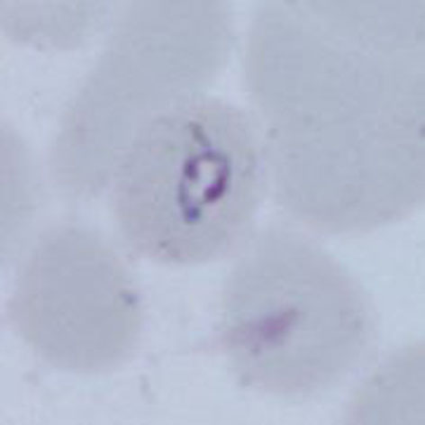
In some cases more than one parasite (most often early or late trophozoites) can be seen within a single erythrocyte. | |||
<gallery mode="nolines" widths=250px heights=250px> | |||
File:double1.jpg|link={{filepath:double1.jpg}} | |||
</gallery> | |||
Note how the chromatin dot of the ring form is divided into two purple masses | |||
<br clear=all> | |||
---- | |||
<span style="color:navy>'''Species significance '''</span> | <span style="color:navy>'''Species significance'''</span> | ||
Most often considered a feature indicating ''P.falciparum'' infection, and is certainly frequent in | Most often considered a feature indicating ''P.falciparum'' infection, and is certainly frequent in that species where it can be used to support the diagnosis. However, the form should not considered as specific, and may occur in any species (and is also a frequent finding for babesia parasites). | ||
---- | ---- | ||
| Line 16: | Line 24: | ||
<span style="color:navy>'''Additional images'''</span> | <span style="color:navy>'''Additional images'''</span> | ||
<gallery mode="nolines" widths=200px heights=200px> | |||
File:double2.jpg|A|link={{filepath:double2.jpg}} | |||
File:double3.jpg|B|link={{filepath:double3.jpg}} | |||
File:double4.jpg|C|link={{filepath:double4.jpg}} | |||
</gallery> | |||
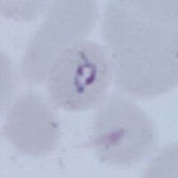
Malaria double chromatin dot forms in: late trophozoite of ''P.ovale'' (A) an early trophozoite of ''P.vivax'' (B) amd eraly trophozoite of P.knowlesi (C) | |||
---- | ---- | ||
Revision as of 19:34, 15 March 2024
What are double infected cells?
In some cases more than one parasite (most often early or late trophozoites) can be seen within a single erythrocyte.
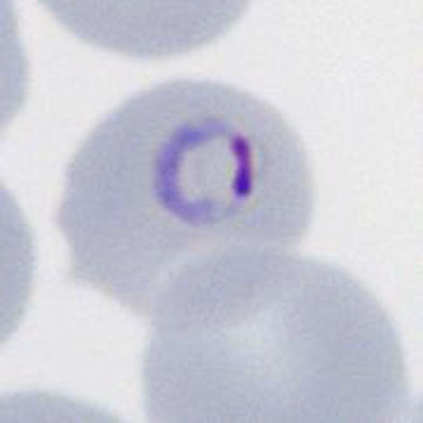
Note how the chromatin dot of the ring form is divided into two purple masses
Species significance
Most often considered a feature indicating P.falciparum infection, and is certainly frequent in that species where it can be used to support the diagnosis. However, the form should not considered as specific, and may occur in any species (and is also a frequent finding for babesia parasites).
Additional images

A

B
C
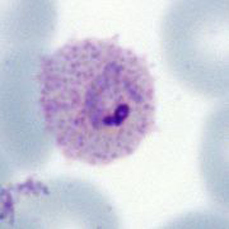
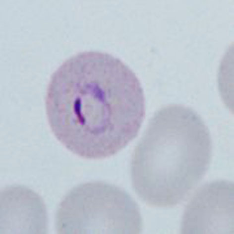
Malaria double chromatin dot forms in: late trophozoite of P.ovale (A) an early trophozoite of P.vivax (B) amd eraly trophozoite of P.knowlesi (C)