Maurer's dots and clefts: Difference between revisions
From haematologyetc.co.uk
(Created page with "Maurer maurer's dots and clefts Description: These are blue-grey dots or linear structures contained within erythrocytes infected by the late trophozoites of P.falciparum and represent modification of the red cell by the parasite. The structures differ from the dot in p.vivax or p.ovale since they are few in number you could imagine being able to count them (in the unlikely case that you wanted to). They are not seen in early trophozoites. Species significance: T...") |
No edit summary |
||
| Line 1: | Line 1: | ||
---- | |||
'''Navigation'''</br> | |||
[[Plasmodium falciparum: Morphology|Go Back]] | |||
---- | |||
{| class="wikitable" style="border-style: solid; border-width: 4px; color:black" | |||
|colspan="1" style = "font-size:100%; color:black; background: FFFAFA"|<span style="color:navy>'''What are double infected cells?'''</span> | |||
These are blue-grey dots or linear structures seen at the late trophozoites of ''P.falciparum''. These structures lie within the erythrocyte cytoplasm and are the result of modification of the red cell by the parasite. The structures differ from the dots seen in ethrocytes infected by ''P.vivax'' or ''P.ovale'' since they are few in number (you could imagine being able to count them). They are not seen in early trophozoites so define the late trophozoite stage. | |||
<gallery mode="nolines" widths=250px heights=250px> | |||
File:11multiple1.jpg|link={{filepath:11multiple1.jpg}} | |||
</gallery> | |||
<span style="font-size:80%">The most frequent form - two early trophozoites of ''P.falciparum'' in a single erythrocyte</span> | |||
<br clear=all> | |||
---- | |||
<span style="color:navy>'''Species significance'''</span> | |||
Maurer's dots and clefts are restricted to ''P.falciparum'' and can be distinguished from Schüffner's dots of ''P.vivax'' or the James' dots of ''P.ovale'' by their blue-grey colour, higher density and sometimes elongated (clefted) shape. | |||
---- | |||
<span style="color:navy>'''Compare with:'''</span> | |||
<gallery mode="nolines" widths=200px heights=200px> | |||
File:11multiple2.jpg|A|link={{filepath:11multiple2.jpg}} | |||
File:11multiple3.jpg|B|link={{filepath:11multiple3.jpg}} | |||
File:11multiple4.jpg|C|link={{filepath:11multiple4.jpg}} | |||
</gallery> | |||
<span style="font-size:80%">Maurer's dots and clefts in a late trophozoite of ''P.falciparum'' (A). These are more dense and blue grey than the softer amd more red Schüffner's dots seen in ''P.vivax'' and James' dots in ''P.ovale'' (C)</span> | |||
Revision as of 13:12, 24 March 2024
Navigation
Go Back
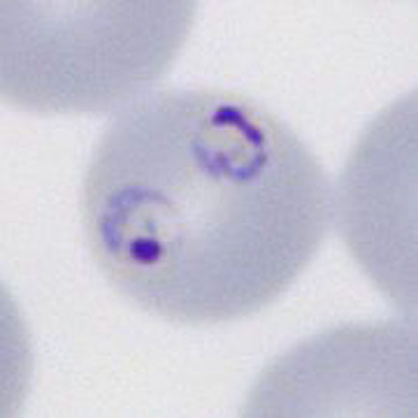
| What are double infected cells?
These are blue-grey dots or linear structures seen at the late trophozoites of P.falciparum. These structures lie within the erythrocyte cytoplasm and are the result of modification of the red cell by the parasite. The structures differ from the dots seen in ethrocytes infected by P.vivax or P.ovale since they are few in number (you could imagine being able to count them). They are not seen in early trophozoites so define the late trophozoite stage.
The most frequent form - two early trophozoites of P.falciparum in a single erythrocyte
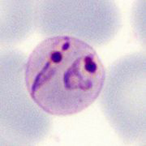
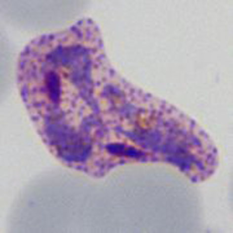
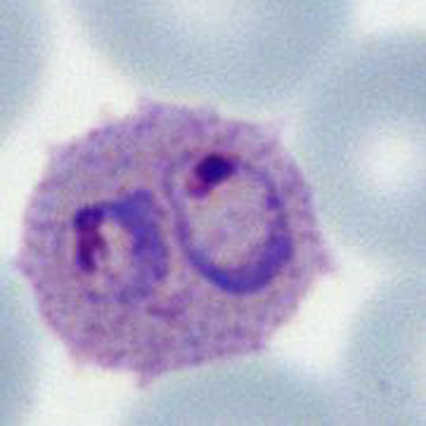
Species significance Maurer's dots and clefts are restricted to P.falciparum and can be distinguished from Schüffner's dots of P.vivax or the James' dots of P.ovale by their blue-grey colour, higher density and sometimes elongated (clefted) shape. Compare with:
Maurer's dots and clefts in a late trophozoite of P.falciparum (A). These are more dense and blue grey than the softer amd more red Schüffner's dots seen in P.vivax and James' dots in P.ovale (C) |