Malaria pigment: Difference between revisions
From haematologyetc.co.uk
No edit summary |
No edit summary |
||
| (7 intermediate revisions by the same user not shown) | |||
| Line 9: | Line 9: | ||
<gallery mode="nolines" widths="220px" heights="220px" > | <gallery mode="nolines" widths="220px" heights="220px" > | ||
File: | File:Pig1.jpg|link={{filepath:MPi1.jpg}} | ||
</gallery> | </gallery> | ||
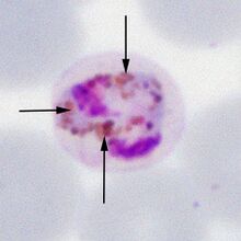
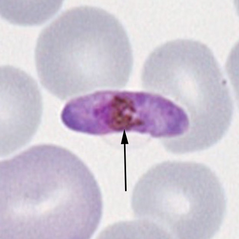
A solid and angular late trophozoite form of ''P.malariae''. Note the golden pigment in separate clumps of granules distributed over the parasite surface (arrowed). | |||
| Line 16: | Line 18: | ||
During their development malarial parasites metabolise the haemoglobin within erythrocytes to support their growth | During their development malarial parasites metabolise the haemoglobin within erythrocytes to support their growth. Evetually infected cells at late stages of parasite development red cell may have no visible haemoglobin. As part of that process the parasite must "detoxify" the iron component of the haem element. This process creates a detoxified iron containing protein "haemazoin" which is visible as pigment - as you might expect this is most visible at late stages of parasite development. | ||
| Line 34: | Line 36: | ||
<gallery mode="nolines" widths="220px" heights="220px" > | <gallery mode="nolines" widths="220px" heights="220px" > | ||
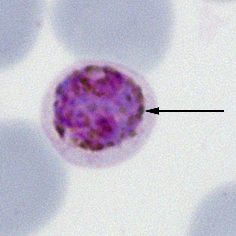
File: | File:Pig2.jpg|Gametocyte ''P.malariae''|link={{filepath:Pig2.jpg}} | ||
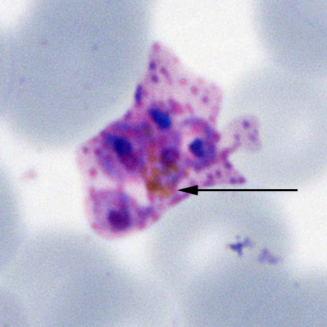
File: | File:Pig3.jpg|Schizont ''P.ovale''|link={{filepath:Pig3.jpg}} | ||
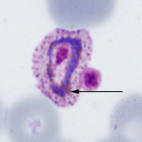
File: | File:Pig4.jpg|Trophozoite ''P.ovale''|link={{filepath:Pig4.jpg}} | ||
File:Pig5.jpg|Gametocyte ''P.falciparum''|link={{filepath:Pig5.jpg}} | |||
</gallery> | </gallery> | ||
Latest revision as of 15:26, 28 March 2024
Navigation
Go Back
| What is malaria pigment?
A solid and angular late trophozoite form of P.malariae. Note the golden pigment in separate clumps of granules distributed over the parasite surface (arrowed).
Pigment may vary in colour and may be clumped or scattered as individual small masses depending on species; in some instances this can help (most obviously in the central clump seen in the "daisy head" schizonts of P.malariae). Generally however, the form of the pigment is less useful than other features in determining species.
Pigment in different stages of parasite development:
|