| Changes to gametocyte morphology in stored blood
NOTE These changes are only observed if blood film preparation is delayed - these are instructive for the changes that occur within the mosquito, and are included as occasionally these may be encountered in laboratory practice and can cause confusion.
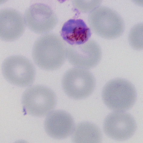
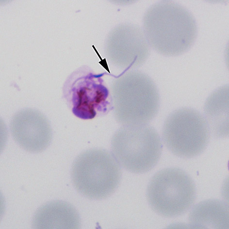
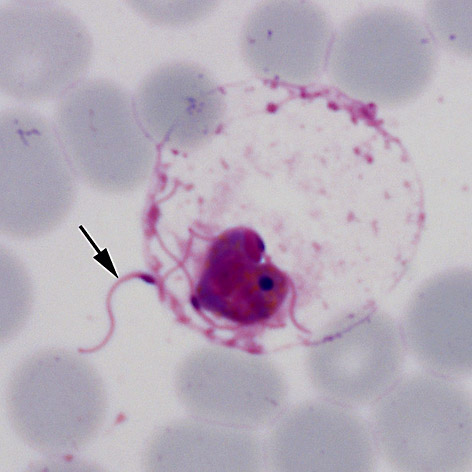
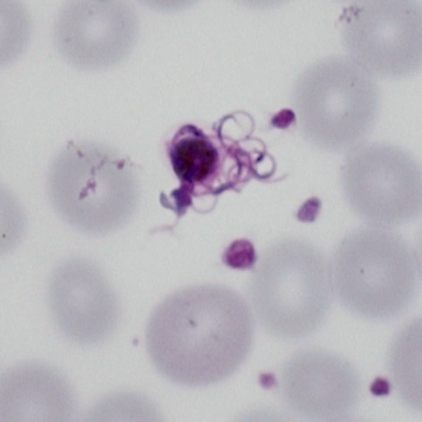
To an extent these changes resemble those that take place within the mosquito midgut. The initial stage involves both the male microgametocytes and female macrogametocytes swelling and becoming more globular. The stage shown in (A) may represent an oocyte although it is not certain (these often clump together). The following stages are quite recognisable for the male gametocte as the male gametes burst from the erythrocytes in the process of exflagellation this may be seen as the early emergence (B), red cell swelling and disollution (C) and emergence of the gametes (D).
|