Haemoglobin crystals: Difference between revisions
From haematologyetc.co.uk
(Created page with " '''Derivation:''' ''Descriptive term: haemoglobin in crystalline form within an erythrocyte'' ---- '''Appearance''' Variously described as tetragonal, hexagonal or rhomboidal crystals, but in practice highly variable shapes resembling rods, solid lumps or geometric shapes (see pathogenesis). The remainder of the cell will have reduced haemoglobin content and may have an empty (“ghost”) appearance. <span style="font-size:90%"><span style="color:#808080"> <gal...") |
No edit summary Tag: Manual revert |
||
| (12 intermediate revisions by the same user not shown) | |||
| Line 10: | Line 10: | ||
Variously described as tetragonal, hexagonal or rhomboidal crystals, but in practice highly variable shapes resembling rods, solid lumps or geometric shapes (see pathogenesis). The remainder of the cell will have reduced haemoglobin content and may have an empty (“ghost”) appearance. | Variously described as tetragonal, hexagonal or rhomboidal crystals, but in practice highly variable shapes resembling rods, solid lumps or geometric shapes (see pathogenesis). The remainder of the cell will have reduced haemoglobin content and may have an empty (“ghost”) appearance. | ||
<gallery mode="nolines" widths="240px" heights="240px" border="1px" > | |||
< | File:Crys0.png|link={{filepath:Crys1.png}} | ||
File:Crys2B.jpg|link={{filepath:Crys2.jpg}} | |||
File: | |||
File: | |||
</gallery> | </gallery> | ||
<span style="font-size:90% | <span style="font-style:italic; font-size:90%> The crystals appear as very dense haemoglobin areas that are classically hexagonal or rhomboidal, but in practice irregular shapes and particularly rods are common. The red cell will often be hypochromic and may appear as a ghost cell as the haemoglobin is condensed into the crystal form. The crystal form of haemoglobin indicates the presence of excess haemoglobin C (HbCC, HbSC, HbC-beta thalassaemia) so look for evidence of other typical cell types for the disorder. | ||
</span> | </span> | ||
| Line 50: | Line 43: | ||
{| class="wikitable" style="color: | <div style="width: 95%; overflow: auto; border: 1px solid navy; font-size:100%"> | ||
{| class="wikitable" style="color:black; background-color:#ffffff;" cellpadding="15" | |||
!colspan="1" |'''The presence of haemoglobin C''' | !colspan="1" |'''The presence of haemoglobin C''' | ||
|- | |- | ||
| Line 57: | Line 51: | ||
|Not HbC trait | |Not HbC trait | ||
|} | |} | ||
</div> | |||
| Line 66: | Line 60: | ||
<gallery widths="250px" heights="250px" > | |||
<gallery widths=" | File:Crys3.jpg|link={{filepath:Crys3.jpg}} | ||
File: | |||
</gallery> | </gallery> | ||
<span style="font-style:italic; font-size:90%;'' > | |||
'''Clinical Image 1''':The most obvious example has two rounded crystals within an empty “ghost” erythrocyte. The surrounding cells are dominated by contracted forms and target cells – some of which have rather dense and square looking “bulls eyes” (bottom centre of image). Clinical condition HbCC | |||
<span style="font-size:90% | <gallery widths="250px" heights="250px" > | ||
File:Crys4.jpg|link={{filepath:Crys4.jpg}} | |||
</gallery> | |||
<span style="font-style:italic; font-size:90%;'' > | |||
''''Clinical Image 2:''' The clearest crystal present is in an elongated cell that has a boat-like form but containing a long central “rod” shaped crystal. Note also the elongated cell (upper left) in which the haemoglobin is contratcted to the two opposing poles of the cell. Clinical condition HbCC | ''''Clinical Image 2:''' The clearest crystal present is in an elongated cell that has a boat-like form but containing a long central “rod” shaped crystal. Note also the elongated cell (upper left) in which the haemoglobin is contratcted to the two opposing poles of the cell. Clinical condition HbCC | ||
<span style="font-size:90% | |||
<gallery widths="250px" heights="250px" > | |||
File:Crys6.jpg|link={{filepath:Crys6.jpg}} | |||
</gallery> | |||
<span style="font-style:italic; font-size:90%;'' > | |||
'''Clinical Image 3:''' A further elongated boat-shaped cell with a central “rod”. Clinical condition HbCC | '''Clinical Image 3:''' A further elongated boat-shaped cell with a central “rod”. Clinical condition HbCC | ||
<span style="font-size:90% | |||
<gallery widths="250px" heights="250px" > | |||
File:Crys5.jpg|link={{filepath:Crys5.jpg}} | |||
</gallery> | |||
<span style="font-style:italic; font-size:90%;'' > | |||
'''Clinical Image 4:''' A ghost (empty) erythrocyte has a single condensed crystal of haemoglobin within it (lower centre of film). A second erythrocyte (lower right quadrant) has a central square of condensed haemoglobin crystal. Clinical condition HbCC | '''Clinical Image 4:''' A ghost (empty) erythrocyte has a single condensed crystal of haemoglobin within it (lower centre of film). A second erythrocyte (lower right quadrant) has a central square of condensed haemoglobin crystal. Clinical condition HbCC | ||
Latest revision as of 17:46, 27 March 2023
Derivation: Descriptive term: haemoglobin in crystalline form within an erythrocyte
Appearance
Variously described as tetragonal, hexagonal or rhomboidal crystals, but in practice highly variable shapes resembling rods, solid lumps or geometric shapes (see pathogenesis). The remainder of the cell will have reduced haemoglobin content and may have an empty (“ghost”) appearance.
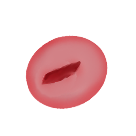
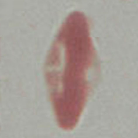
The crystals appear as very dense haemoglobin areas that are classically hexagonal or rhomboidal, but in practice irregular shapes and particularly rods are common. The red cell will often be hypochromic and may appear as a ghost cell as the haemoglobin is condensed into the crystal form. The crystal form of haemoglobin indicates the presence of excess haemoglobin C (HbCC, HbSC, HbC-beta thalassaemia) so look for evidence of other typical cell types for the disorder.
Significance
The crystallisation process results from altered electrostatic properties of HbC caused by the loss of charge associated with the mutation– crystals therefore imply the presence of HbC. They form in homozygous HbC disease (HbCC), but may be found in HbSC and HbC/beta thalassaemia (they do not form in HbC heterozygotes).
Pathobiology
In Hb C, glutamic acid in the sixth position of the β-globin chain is replaced by lysine. This amino acid substitution alters the electrostatic interactions of the haemoglobin molecule leading to decreased solubility in its de-oxygenated (or hypertonic/dehydrated) state causing crystal formation. Impurities (other red cell proteins and denatured haemoglobin) are incorporated into the crystals and severely affect their growth so that perfect crystals tend not to formed: although often described as hexagonal or rhomboidal, very irregular or rod-like crystals are often seen.
Pitfalls
The crystal formation will often involve a large portion of the intracellular haemoglobin leaving an empty membrane shell. For this reason, there may be confusion with a hemighost or blister cell. The key here is to recognise the cellular background (particularly frequent target cells or boat cells), but also to appreciate the appearance of crystals.
Causes
| The presence of haemoglobin C |
|---|
| Signifies any cell where HbC is present in significant amount: HbCC, HbSC HbC-beta thal. |
| Not HbC trait |
Clinical Examples
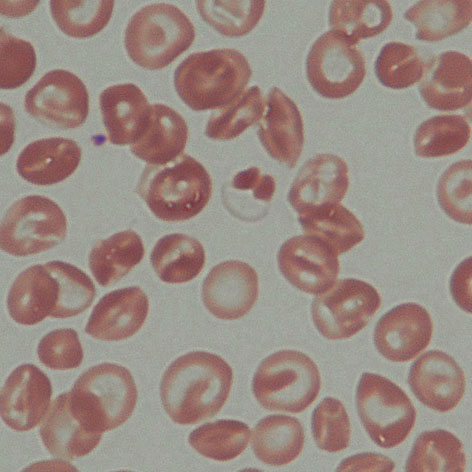
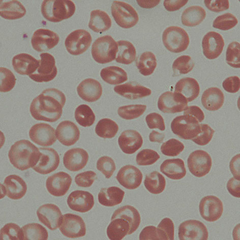
Clinical Image 1:The most obvious example has two rounded crystals within an empty “ghost” erythrocyte. The surrounding cells are dominated by contracted forms and target cells – some of which have rather dense and square looking “bulls eyes” (bottom centre of image). Clinical condition HbCC
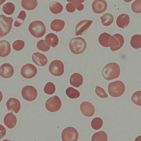
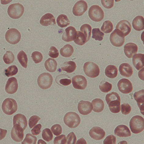
'Clinical Image 2: The clearest crystal present is in an elongated cell that has a boat-like form but containing a long central “rod” shaped crystal. Note also the elongated cell (upper left) in which the haemoglobin is contratcted to the two opposing poles of the cell. Clinical condition HbCC
Clinical Image 3: A further elongated boat-shaped cell with a central “rod”. Clinical condition HbCC
Clinical Image 4: A ghost (empty) erythrocyte has a single condensed crystal of haemoglobin within it (lower centre of film). A second erythrocyte (lower right quadrant) has a central square of condensed haemoglobin crystal. Clinical condition HbCC