Click for images of schizont morphology: Difference between revisions
From haematologyetc.co.uk
(Created page with "---- '''Navigation'''</br> Go Back ---- {| class="wikitable" style="border-style: solid; border-width: 4px; color:black" |colspan="1" style = "font-size:100%; color:black; background: FFFAFA"|<span style="color:navy>'''How does schizont appearance change during their development?'''</span> Schizonts formation involves successive cycles of asexual division that eventually result in the formation of multiple separate "merozoite" fo...") |
No edit summary |
||
| Line 1: | Line 1: | ||
---- | ---- | ||
'''Navigation'''</br> | '''Navigation'''</br> | ||
[[ | [[Biology of the schizont|Go Back]] | ||
---- | ---- | ||
Latest revision as of 18:24, 17 April 2024
Navigation
Go Back
| How does schizont appearance change during their development?
THE INITIAL ASEXUAL DIVISION
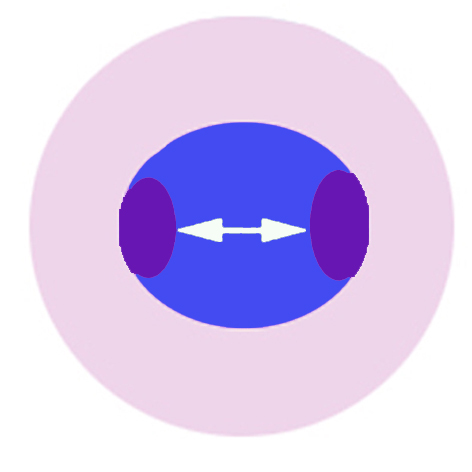
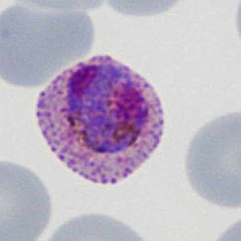
The cartoon image (A) shows the division of chromatin into two distinct purple chromatin masses within the blue parasite cytoplasm (at this point the cytoplams is not divided so indiviual merozoites are not really distinguishable). A clinical image of a parasite at this developmental stage (P.ovale with well shown James'dots) is shown in panel (B).
IMMATURE SCHIZONT APPEARANCES
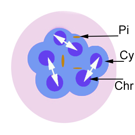
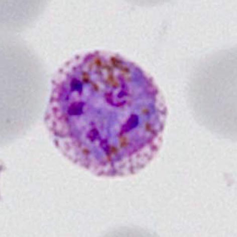
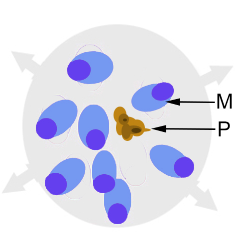
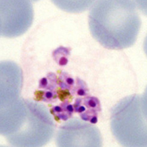
The cartoon image (A) shows the further division of chromatin (Chr) into many discrete massed within the blue parasite cytoplasm (Cy). Indiviual merozoites are still not distinguishable but the malaria pigment is obvious (Pi). A clinical image of a parasite at this developmental stage (again from P.ovale with well shown James'dots and malaria pigment) is shown in panel (B).
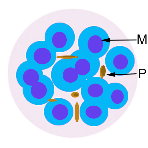
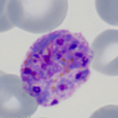
MATURE SCHIZONT APPEARANCES By this stage the individual merozoites can be distinguished, each with a chromatin dot and cytoplasm; they are now ready for release from the red cell.
In the final stage the red cell membrane is broken down, swelling then separating to release the merozoites and any malaria pigment into the blood where each merozoite enters a red cell to form a new early trophozoite and increasing the infection load.
|