Accolé form: Difference between revisions
From haematologyetc.co.uk
(Created page with "---- '''Description''' Accolé forms may also be referred to as "edge" or “appliqué” form, and describes parasites in early or late trophozoites where the parasite appears closely opposed to the edge of the erythrocyte membrane - usually appearing to be flattened against the erythrocyte membrane. IMAGE '''Species significance''' Most often considered to be a feature of p.falciparum infection and can be helpful to indicate this diagnosis (particularly when freq...") |
No edit summary |
||
| Line 1: | Line 1: | ||
---- | |||
'''Navigation'''</br> | |||
[[Plasmodium falciparum: Morphology|Go Back]] | |||
---- | ---- | ||
''' | {| class="wikitable" style="border-style: solid; border-width: 5px; color:black" | ||
|colspan="1" style = "font-size:100%; color:black; background: WhiteSmoke"|<span style="color:navy>'''What is a double dot form?'''</span> | |||
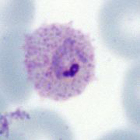
Accolé forms may also be referred to as "edge" or “appliqué” form, and describes parasites in early or late trophozoites where the parasite appears closely opposed to the edge of the erythrocyte membrane - usually appearing to be flattened against the erythrocyte membrane. | Accolé forms may also be referred to as "edge" or “appliqué” form, and describes parasites in early or late trophozoites where the parasite appears closely opposed to the edge of the erythrocyte membrane - usually appearing to be flattened against the erythrocyte membrane. | ||
'' | <gallery mode="nolines" widths=250px heights=250px> | ||
File:PFaccole.jpg|link={{filepath:PFaccole.jpg}} | |||
</gallery> | |||
<span style="font-size:80%">Note that the parasite is very closely in contact with the red cell membrane (''P.falciparum'' late trophozoite form with maurer's dots and clefts</span> | |||
<br clear=all> | |||
Most often considered to be a feature of p.falciparum infection and | ---- | ||
<span style="color:navy>'''Species significance'''</span> | |||
Most often considered to be a feature of p.falciparum infection and when frequent these appearances are helpful to indicate this species. However, the form is not fully specific and examples may occur in any species. | |||
---- | ---- | ||
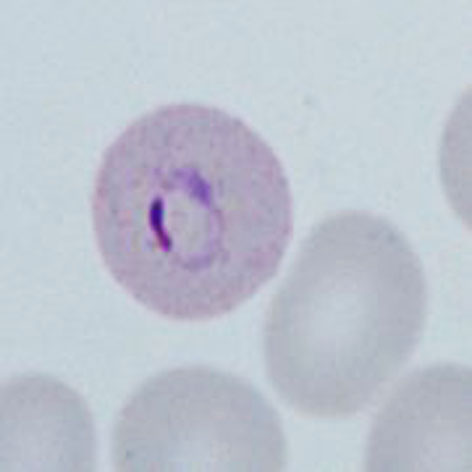
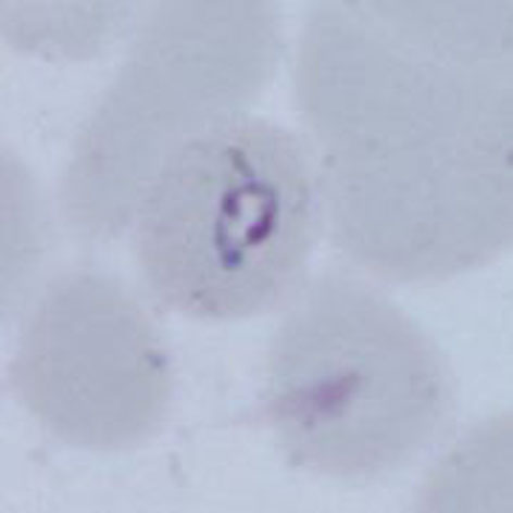
'''Additional | <span style="color:navy>'''Additional images'''</span> | ||
<gallery mode="nolines" widths=200px heights=200px> | |||
File:double2.jpg|A|link={{filepath:double2.jpg}} | |||
File:double3.jpg|B|link={{filepath:double3.jpg}} | |||
File:double4.jpg|C|link={{filepath:double4.jpg}} | |||
</gallery> | |||
<span style="font-size:80%">Malaria double chromatin dot forms in: late trophozoite of ''P.ovale'' (A) an early trophozoite of ''P.vivax'' (B) and ealy trophozoite of P.knowlesi (C)<span> | |||
---- | ---- | ||
Revision as of 13:08, 19 March 2024
Navigation
Go Back
| What is a double dot form?
Accolé forms may also be referred to as "edge" or “appliqué” form, and describes parasites in early or late trophozoites where the parasite appears closely opposed to the edge of the erythrocyte membrane - usually appearing to be flattened against the erythrocyte membrane.
Note that the parasite is very closely in contact with the red cell membrane (P.falciparum late trophozoite form with maurer's dots and clefts
Species significance Most often considered to be a feature of p.falciparum infection and when frequent these appearances are helpful to indicate this species. However, the form is not fully specific and examples may occur in any species. Additional images
Malaria double chromatin dot forms in: late trophozoite of P.ovale (A) an early trophozoite of P.vivax (B) and ealy trophozoite of P.knowlesi (C) |