Plasmodium vivax: Morphology: Difference between revisions
From haematologyetc.co.uk
No edit summary |
No edit summary |
||
| Line 51: | Line 51: | ||
The later growth stage: | The later growth stage: | ||
* | *infected erythrocytes become significantly enlarged and irregular in shape | ||
* | *parasites lose their ring appearnace becoming irregular and "amoeboid" in form | ||
*numerous red/purple Schuffners dots are predent in the cytoplasm of red cells | |||
*malaria pigment is often present and has an irregular distribution | |||
* | |||
| Line 130: | Line 129: | ||
the schizont file pvs.jpg leftt 200px link filepath pvs.jpg *a range of maturing schizonts will generally be present within enlarged red cells *mature schizonts generally contain 16-24 separate merozoites *schu8c3bcffneru8e28099s dots can be detected in any residual cytoplasm of the erythrocyte *pigment is visible in irregularly distributed clumps over the schizont surface | the schizont file pvs.jpg leftt 200px link filepath pvs.jpg *a range of maturing schizonts will generally be present within enlarged red cells *mature schizonts generally contain 16-24 separate merozoites *schu8c3bcffneru8e28099s dots can be detected in any residual cytoplasm of the erythrocyte *pigment is visible in irregularly distributed clumps over the schizont surface | ||
the gametocyte file pvg.jpg leftt 200px link filepath pvg.jpg *very large with ovoid or distorted forms *macrogametocytes female may entirely fill the erythrocyte *microgametocytes male may have a thin cytoplasmic rim with visible schu8c3bcffneru8e28099s dots *pigment is clumped over the surface of the gametocyte | the gametocyte file pvg.jpg leftt 200px link filepath pvg.jpg *very large with ovoid or distorted forms *macrogametocytes female may entirely fill the erythrocyte *microgametocytes male may have a thin cytoplasmic rim with visible schu8c3bcffneru8e28099s dots *pigment is clumped over the surface of the gametocyte | ||
Revision as of 23:37, 3 April 2024
Navigation
(click blue highlighted text to return to page)
Malaria main index
>Species identification: summary page
>>This page: P.vivax: morphology
| The early trophozoite |
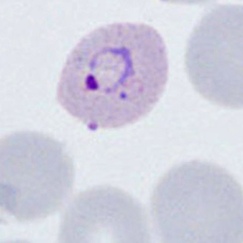
The earliest ring forms may be indistinguishable from other species, but during this stage the parasite tends to aquire a more irregular forms and to show signs of modification of the erythrocyte (added dots, and altered size and shape).
- erythrocytes begin to show increased size and altered shape
- parasites retain a ring form but may aquire a more irregular form
- parasites are generally large - occupying up to half of the erythrocyte
- cytoplasmic Schüffner's dots may appear at this stage, although pigment is less uncommon
| The late trophozoite |
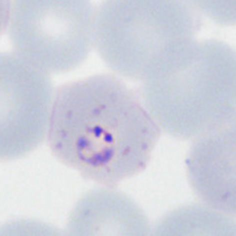
The later growth stage:
- infected erythrocytes become significantly enlarged and irregular in shape
- parasites lose their ring appearnace becoming irregular and "amoeboid" in form
- numerous red/purple Schuffners dots are predent in the cytoplasm of red cells
- malaria pigment is often present and has an irregular distribution
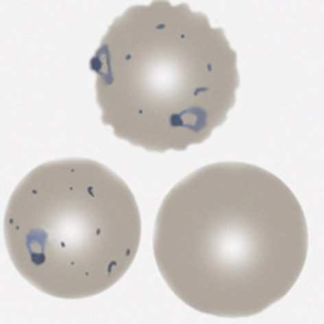
| The schizont |
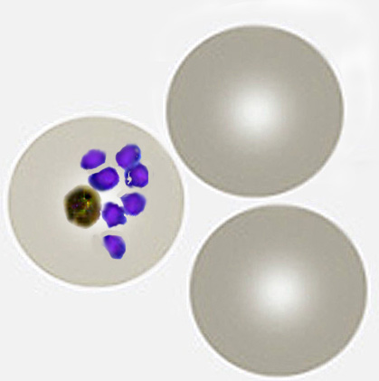
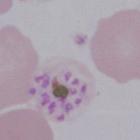
The asexual form:
- Do not generally circulate in this species unless overwhelming infection
- The asexually formed developing "merozoites" cluster untidily
- Schizonts develop progressively to form 8-16 merozoites when mature
- In this species the loose malaria pigment may be seen in clumps between the parasites
- Red cell size is generally unaffected but red cells become pale as haemoglobin is metabolised by the parasites
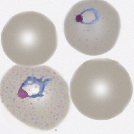
The gametocyte
| The gametocyte |
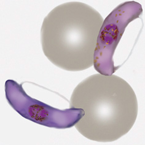
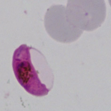
The sexual replication form (very distinctive).
- Gametocytes are elongated but are restricted into typical shape by the red cell membrane
- They parasites are rod shaped but the membrane may cause them to curve into a “"banana" form”
- The residual membrane (empty of haemoglobin) is often seen as a "blister" to the side of the parasite
- The single chromatin area is in the centre of the parasite, often has pigment overlying it
- Gametocytes may not be be seen, or may be the only form present (particularly after treatment)
the schizont file pvs.jpg leftt 200px link filepath pvs.jpg *a range of maturing schizonts will generally be present within enlarged red cells *mature schizonts generally contain 16-24 separate merozoites *schu8c3bcffneru8e28099s dots can be detected in any residual cytoplasm of the erythrocyte *pigment is visible in irregularly distributed clumps over the schizont surface
the gametocyte file pvg.jpg leftt 200px link filepath pvg.jpg *very large with ovoid or distorted forms *macrogametocytes female may entirely fill the erythrocyte *microgametocytes male may have a thin cytoplasmic rim with visible schu8c3bcffneru8e28099s dots *pigment is clumped over the surface of the gametocyte